 Movie
Movie Controller


+ Open data

- Basic information
| Entry | Database: PDB / ID: 5jul | |||||||||||||||||||||||||||||||||||||||||||||
|---|---|---|---|---|---|---|---|---|---|---|---|---|---|---|---|---|---|---|---|---|---|---|---|---|---|---|---|---|---|---|---|---|---|---|---|---|---|---|---|---|---|---|---|---|---|---|
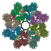
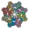
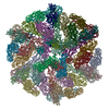
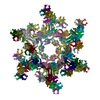
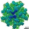
| Title | Near atomic structure of the Dark apoptosome | |||||||||||||||||||||||||||||||||||||||||||||
 Components Components | Apaf-1 related killer DARK | |||||||||||||||||||||||||||||||||||||||||||||
 Keywords Keywords | APOPTOSIS / Dark / apoptosome / apotosis / AAA+ ATPase | |||||||||||||||||||||||||||||||||||||||||||||
| Function / homology |  Function and homology information Function and homology informationnegative regulation of humoral immune response / positive regulation of glial cell apoptotic process / Formation of apoptosome / salivary gland histolysis / melanization defense response / : / Regulation of the apoptosome activity / Activation of caspases through apoptosome-mediated cleavage / compound eye retinal cell programmed cell death / central nervous system formation ...negative regulation of humoral immune response / positive regulation of glial cell apoptotic process / Formation of apoptosome / salivary gland histolysis / melanization defense response / : / Regulation of the apoptosome activity / Activation of caspases through apoptosome-mediated cleavage / compound eye retinal cell programmed cell death / central nervous system formation / positive regulation of apoptotic process involved in development / sperm individualization / apoptosome / autophagic cell death / chaeta development / Neutrophil degranulation / L-methionine cycle / CARD domain binding / programmed cell death / triglyceride homeostasis / dendrite morphogenesis / response to starvation / response to gamma radiation / neuron cellular homeostasis / ADP binding / apoptotic process / structural molecule activity / ATP binding / identical protein binding Similarity search - Function | |||||||||||||||||||||||||||||||||||||||||||||
| Biological species |  | |||||||||||||||||||||||||||||||||||||||||||||
| Method | ELECTRON MICROSCOPY / single particle reconstruction / cryo EM / Resolution: 4.4 Å | |||||||||||||||||||||||||||||||||||||||||||||
 Authors Authors | Cheng, T.C. / Akey, I.V. / Yuan, S. / Yu, Z. / Ludtke, S.J. / Akey, C.W. | |||||||||||||||||||||||||||||||||||||||||||||
| Funding support |  United States, 1items United States, 1items
| |||||||||||||||||||||||||||||||||||||||||||||
 Citation Citation | Journal: Structure / Year: 2017 Title: A Near-Atomic Structure of the Dark Apoptosome Provides Insight into Assembly and Activation. Authors: Tat Cheung Cheng / Ildikó V Akey / Shujun Yuan / Zhiheng Yu / Steven J Ludtke / Christopher W Akey / Abstract: In Drosophila, the Apaf-1-related killer (Dark) forms an apoptosome that activates procaspases. To investigate function, we have determined a near-atomic structure of Dark double rings using cryo- ...In Drosophila, the Apaf-1-related killer (Dark) forms an apoptosome that activates procaspases. To investigate function, we have determined a near-atomic structure of Dark double rings using cryo-electron microscopy. We then built a nearly complete model of the apoptosome that includes 7- and 8-blade β-propellers. We find that the preference for dATP during Dark assembly may be governed by Ser325, which is in close proximity to the 2' carbon of the deoxyribose ring. Interestingly, β-propellers in V-shaped domains of the Dark apoptosome are more widely separated, relative to these features in the Apaf-1 apoptosome. This wider spacing may be responsible for the lack of cytochrome c binding to β-propellers in the Dark apoptosome. Our structure also highlights the roles of two loss-of-function mutations that may block Dark assembly. Finally, the improved model provides a framework to understand apical procaspase activation in the intrinsic cell death pathway. | |||||||||||||||||||||||||||||||||||||||||||||
| History |
|
- Structure visualization
| Movie |
Movie viewer |
|---|---|
| Structure viewer | Molecule: MolmilJmol/JSmol |
- Downloads & links
-Download
| PDBx/mmCIF format | 5jul.cif.gz | 3.3 MB | Display | PDBx/mmCIF format |
|---|---|---|---|---|
| PDB format | pdb5jul.ent.gz | Display | PDB format | |
| PDBx/mmJSON format | 5jul.json.gz | Tree view | PDBx/mmJSON format | |
| Others |  Other downloads Other downloads |
-Validation report
| Arichive directory | https://data.pdbj.org/pub/pdb/validation_reports/ju/5julftp://data.pdbj.org/pub/pdb/validation_reports/ju/5jul | HTTPS FTP |
|---|
-Related structure data
| Related structure data |  8177MC M: map data used to model this data C: citing same article ( |
|---|---|
| Similar structure data |

-Links
 PDBj
PDBj


- Assembly
| Deposited unit | 
|
|---|---|
| 1 |
|
-Components
| #1: Protein | Mass: 166131.000 Da / Num. of mol.: 16 Source method: isolated from a genetically manipulated source Source: (gene. exp.) Gene: Dark, Ark, Ark-RB, dapaf-1L, Hac1, CG6829, Dmel_CG6829 Production host:  Spodoptera frugiperda (fall armyworm) / References: UniProt: Q7KLI1 Spodoptera frugiperda (fall armyworm) / References: UniProt: Q7KLI1#2: Chemical | ChemComp-DTP /   Mass: 491.182 Da / Num. of mol.: 16 / Source method: obtained synthetically / Formula: C10H16N5O12P3 Mass: 491.182 Da / Num. of mol.: 16 / Source method: obtained synthetically / Formula: C10H16N5O12P3Has protein modification | Y | |
|---|
-Experimental details
-Experiment
| Experiment | Method: ELECTRON MICROSCOPY |
|---|---|
| EM experiment | Aggregation state: PARTICLE / 3D reconstruction method: single particle reconstruction |
- Sample preparation
| Component | Name: Dark apoptosome / Type: COMPLEX / Entity ID: #1 / Source: RECOMBINANT | ||||||||||||||||||||||||||||||
|---|---|---|---|---|---|---|---|---|---|---|---|---|---|---|---|---|---|---|---|---|---|---|---|---|---|---|---|---|---|---|---|
| Molecular weight | Value: 2.3 MDa / Experimental value: NO | ||||||||||||||||||||||||||||||
| Source (natural) | Organism: | ||||||||||||||||||||||||||||||
| Source (recombinant) | Organism: Spodoptera frugiperda (fall armyworm) / Plasmid: pFastBac | ||||||||||||||||||||||||||||||
| Buffer solution | pH: 7.5 / Details: Buffer A | ||||||||||||||||||||||||||||||
| Buffer component |
| ||||||||||||||||||||||||||||||
| Specimen | Conc.: 2 mg/ml / Embedding applied: NO / Shadowing applied: NO / Staining applied: NO / Vitrification applied: YES | ||||||||||||||||||||||||||||||
| Specimen support | Grid material: COPPER / Grid mesh size: 300 divisions/in. / Grid type: C-flat-1.2/1.3 | ||||||||||||||||||||||||||||||
| Vitrification | Instrument: FEI VITROBOT MARK III / Cryogen name: ETHANE / Humidity: 100 % / Chamber temperature: 293 K Details: 2.5ul sample blot additives present at the indicated final concentrations: NP-40 (0.025%), DHPG (diheptanoylphosphatidylglycerol; 0.05%), cytochrome c (0.5 mg/ml), DeoxybigChaps (0.01%), and lysine (0.03 mg/ml) |
- Electron microscopy imaging
| Experimental equipment |  Model: Titan Krios / Image courtesy: FEI Company |
|---|---|
| Microscopy | Model: FEI TITAN KRIOS |
| Electron gun | Electron source:  FIELD EMISSION GUN / Accelerating voltage: 300 kV / Illumination mode: FLOOD BEAM FIELD EMISSION GUN / Accelerating voltage: 300 kV / Illumination mode: FLOOD BEAM |
| Electron lens | Mode: BRIGHT FIELD / Nominal magnification: 81000 X / Nominal defocus max: 2400 nm / Nominal defocus min: 1500 nm / Cs: 0.01 mm / C2 aperture diameter: 100 µm / Alignment procedure: COMA FREE |
| Specimen holder | Cryogen: NITROGEN / Specimen holder model: FEI TITAN KRIOS AUTOGRID HOLDER |
| Image recording | Average exposure time: 0.3 sec. / Electron dose: 1.8 e/Å2 / Detector mode: COUNTING / Film or detector model: GATAN K2 SUMMIT (4k x 4k) / Num. of grids imaged: 1 / Num. of real images: 1991 |
| EM imaging optics | Energyfilter name: GIF / Spherical aberration corrector: FEI Cs corrector |
| Image scans | Width: 7476 / Height: 7420 / Movie frames/image: 23 / Used frames/image: 2-23 |
- Processing
| EM software |
| ||||||||||||||||||||||||||||||||||||||||
|---|---|---|---|---|---|---|---|---|---|---|---|---|---|---|---|---|---|---|---|---|---|---|---|---|---|---|---|---|---|---|---|---|---|---|---|---|---|---|---|---|---|
| CTF correction | Type: PHASE FLIPPING AND AMPLITUDE CORRECTION | ||||||||||||||||||||||||||||||||||||||||
| Particle selection | Num. of particles selected: 88485 | ||||||||||||||||||||||||||||||||||||||||
| Symmetry | Point symmetry: D8 (2x8 fold dihedral) | ||||||||||||||||||||||||||||||||||||||||
| 3D reconstruction | Resolution: 4.4 Å / Resolution method: FSC 0.143 CUT-OFF / Num. of particles: 17769 / Algorithm: FOURIER SPACE / Symmetry type: POINT | ||||||||||||||||||||||||||||||||||||||||
| Atomic model building | Protocol: FLEXIBLE FIT Details: local fitting with Chimera followed by flexible fitting with MDFF and refinement with Phenix. |
