 Movie
Movie Controller

[English] 日本語
 Yorodumi
- PDB-4v96: The structure of a 1.8 MDa viral genome injection device suggests... -
+ Open data

- Basic information
| Entry | Database: PDB / ID: 4v96 | |||||||||
|---|---|---|---|---|---|---|---|---|---|---|
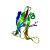
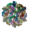
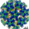
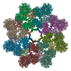
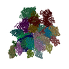
| Title | The structure of a 1.8 MDa viral genome injection device suggests alternative infection mechanisms | |||||||||
 Components Components |
| |||||||||
 Keywords Keywords | VIRAL PROTEIN / Distal tail protein / Receptor-binding protein / Phage baseplate / host adsorption apparatus / genome injection device | |||||||||
| Function / homology |  Function and homology information Function and homology informationvirus tail, baseplate / cell adhesion / symbiont entry into host cell / virion attachment to host cell Similarity search - Function | |||||||||
| Biological species |  Lactococcus phage TP901-1 (virus) Lactococcus phage TP901-1 (virus) | |||||||||
| Method |  X-RAY DIFFRACTION / SYNCHROTRON / SAD / Resolution: 3.8 Å X-RAY DIFFRACTION / SYNCHROTRON / SAD / Resolution: 3.8 Å | |||||||||
 Authors Authors | Veesler, D. / Spinelli, S. / Mahony, J. / Lichiere, J. / Blangy, S. / Bricogne, G. / Legrand, P. / Ortiz-Lombardia, M. / Campanacci, V. / van Sinderen, D. / Cambillau, C. | |||||||||
 Citation Citation | Journal: Proc.Natl.Acad.Sci.USA / Year: 2012 Title: Structure of the phage TP901-1 1.8 MDa baseplate suggests an alternative host adhesion mechanism. Authors: Veesler, D. / Spinelli, S. / Mahony, J. / Lichiere, J. / Blangy, S. / Bricogne, G. / Legrand, P. / Ortiz-Lombardia, M. / Campanacci, V. / van Sinderen, D. / Cambillau, C. | |||||||||
| History |
|
- Structure visualization
| Structure viewer | Molecule: MolmilJmol/JSmol |
|---|
- Downloads & links
-Download
| PDBx/mmCIF format | 4v96.cif.gz | 4.1 MB | Display | PDBx/mmCIF format |
|---|---|---|---|---|
| PDB format | pdb4v96.ent.gz | Display | PDB format | |
| PDBx/mmJSON format | 4v96.json.gz | Tree view | PDBx/mmJSON format | |
| Others |  Other downloads Other downloads |
-Validation report
| Arichive directory | https://data.pdbj.org/pub/pdb/validation_reports/v9/4v96ftp://data.pdbj.org/pub/pdb/validation_reports/v9/4v96 | HTTPS FTP |
|---|
-Related structure data


-Links
 PDBj
PDBj
- Assembly
| Deposited unit | 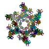
| ||||||||
|---|---|---|---|---|---|---|---|---|---|
| 1 |
| ||||||||
| Unit cell |
|
-Components
| #1: Protein | Mass: 33876.148 Da / Num. of mol.: 18 Source method: isolated from a genetically manipulated source Source: (gene. exp.) Lactococcus phage TP901-1 (virus) / Gene: BppU (ORF48) / Plasmid: pETG20A / Production host:  #2: Protein | Mass: 29085.857 Da / Num. of mol.: 6 Source method: isolated from a genetically manipulated source Source: (gene. exp.) Lactococcus phage TP901-1 (virus) / Gene: Dit (ORF46) / Plasmid: pETG20A / Production host: #3: Protein | Mass: 18365.545 Da / Num. of mol.: 54 Source method: isolated from a genetically manipulated source Source: (gene. exp.) Lactococcus phage TP901-1 (virus) / Gene: bpp, BppL (ORF49) / Plasmid: pETG20A / Production host: |
|---|
-Experimental details
-Experiment
| Experiment | Method: X-RAY DIFFRACTION / Number of used crystals: 1 |
|---|
- Sample preparation
| Crystal | Density Matthews: 4.19 Å3/Da / Density % sol: 70.61 % |
|---|---|
| Crystal grow | Temperature: 293 K / Method: vapor diffusion / pH: 8 Details: 0.08M Trizma pH8.0, 0.1M KCl, 15% PEG2000MME, VAPOR DIFFUSION, temperature 293K |
-Data collection
| Diffraction | Mean temperature: 100 K |
|---|---|
| Diffraction source | Source: SYNCHROTRON / Site: SOLEIL  / Beamline: PROXIMA 1 / Wavelength: 0.8856 Å / Beamline: PROXIMA 1 / Wavelength: 0.8856 Å |
| Detector | Type: ADSC QUANTUM 315r / Detector: CCD / Date: Jul 18, 2010 |
| Radiation | Protocol: SINGLE WAVELENGTH / Monochromatic (M) / Laue (L): M / Scattering type: x-ray |
| Radiation wavelength | Wavelength: 0.8856 Å / Relative weight: 1 |
| Reflection | Resolution: 3.8→45 Å / Num. all: 286540 / Num. obs: 284699 / % possible obs: 99.2 % / Observed criterion σ(I): 1.2 / Redundancy: 3.4 % / Biso Wilson estimate: 106.87 Å2 / Rsym value: 0.108 / Net I/σ(I): 8.72 |
| Reflection shell | Resolution: 3.8→4 Å / Redundancy: 3.15 % / Mean I/σ(I) obs: 1.28 / Num. unique all: 40328 / Rsym value: 0.999 / % possible all: 99.1 |
- Processing
| Software |
| ||||||||||||||||||||||||||||||||||||||||||||||||||||||||||||||||||
|---|---|---|---|---|---|---|---|---|---|---|---|---|---|---|---|---|---|---|---|---|---|---|---|---|---|---|---|---|---|---|---|---|---|---|---|---|---|---|---|---|---|---|---|---|---|---|---|---|---|---|---|---|---|---|---|---|---|---|---|---|---|---|---|---|---|---|---|
| Refinement | Method to determine structure: SAD / Resolution: 3.8→35.97 Å / Cor.coef. Fo:Fc: 0.9148 / Cor.coef. Fo:Fc free: 0.8671 / Isotropic thermal model: isotropic / Cross valid method: THROUGHOUT / σ(F): 0 / Stereochemistry target values: Engh & Huber
| ||||||||||||||||||||||||||||||||||||||||||||||||||||||||||||||||||
| Displacement parameters | Biso mean: 171.32 Å2
| ||||||||||||||||||||||||||||||||||||||||||||||||||||||||||||||||||
| Refine analyze | Luzzati coordinate error obs: 1.291 Å | ||||||||||||||||||||||||||||||||||||||||||||||||||||||||||||||||||
| Refinement step | Cycle: LAST / Resolution: 3.8→35.97 Å
| ||||||||||||||||||||||||||||||||||||||||||||||||||||||||||||||||||
| Refine LS restraints |
| ||||||||||||||||||||||||||||||||||||||||||||||||||||||||||||||||||
| LS refinement shell | Resolution: 3.8→3.9 Å / Total num. of bins used: 20
|
