 Movie
Movie Controller

[English] 日本語
 Yorodumi
- PDB-5jtd: Crystal structure of the Ru(bpy)2PhenA functionalized P450 BM3 L4... -
+ Open data

- Basic information
| Entry | Database: PDB / ID: 5jtd | ||||||
|---|---|---|---|---|---|---|---|
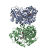
| Title | Crystal structure of the Ru(bpy)2PhenA functionalized P450 BM3 L407C heme domain mutant in complex with DMSO. | ||||||
 Components Components | Bifunctional cytochrome P450/NADPH--P450 reductase | ||||||
 Keywords Keywords | OXIDOREDUCTASE / hybrid P450 BM3 enzymes / electron transfer / photosensitizer / photocatalytic activity | ||||||
| Function / homology |  Function and homology information Function and homology informationaromatase activity / NADPH-hemoprotein reductase / NADPH-hemoprotein reductase activity / oxidoreductase activity, acting on paired donors, with incorporation or reduction of molecular oxygen, reduced flavin or flavoprotein as one donor, and incorporation of one atom of oxygen / unspecific monooxygenase / FMN binding / flavin adenine dinucleotide binding / iron ion binding / heme binding / identical protein binding / cytosol Similarity search - Function | ||||||
| Biological species |  Bacillus megaterium (bacteria) Bacillus megaterium (bacteria) | ||||||
| Method |  X-RAY DIFFRACTION / SYNCHROTRON / MOLECULAR REPLACEMENT / molecular replacement / Resolution: 1.5 Å X-RAY DIFFRACTION / SYNCHROTRON / MOLECULAR REPLACEMENT / molecular replacement / Resolution: 1.5 Å | ||||||
 Authors Authors | Kloos, M. | ||||||
 Citation Citation | Journal: Biochim.Biophys.Acta / Year: 2016 Title: Insights into an efficient light-driven hybrid P450 BM3 enzyme from crystallographic, spectroscopic and biochemical studies. Authors: Spradlin, J. / Lee, D. / Mahadevan, S. / Mahomed, M. / Tang, L. / Lam, Q. / Colbert, A. / Shafaat, O.S. / Goodin, D. / Kloos, M. / Kato, M. / Cheruzel, L.E. | ||||||
| History |
|
- Structure visualization
| Structure viewer | Molecule: MolmilJmol/JSmol |
|---|
- Downloads & links
-Download
| PDBx/mmCIF format | 5jtd.cif.gz | 217.3 KB | Display | PDBx/mmCIF format |
|---|---|---|---|---|
| PDB format | pdb5jtd.ent.gz | 170.8 KB | Display | PDB format |
| PDBx/mmJSON format | 5jtd.json.gz | Tree view | PDBx/mmJSON format | |
| Others |  Other downloads Other downloads |
-Validation report
| Arichive directory | https://data.pdbj.org/pub/pdb/validation_reports/jt/5jtdftp://data.pdbj.org/pub/pdb/validation_reports/jt/5jtd | HTTPS FTP |
|---|
-Related structure data
| Related structure data |  5jq2C  1jpzS S: Starting model for refinement C: citing same article ( |
|---|---|
| Similar structure data |


-Links
 PDBj
PDBj


- Assembly
| Deposited unit | 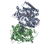
| ||||||||
|---|---|---|---|---|---|---|---|---|---|
| 1 |
| ||||||||
| Unit cell |
|
-Components
| #1: Protein | Mass: 53685.062 Da / Num. of mol.: 2 / Mutation: L407C Source method: isolated from a genetically manipulated source Details: A Ru(bpy)2)PhenA was covalently attached to a non-native single cysteine (L407C) mutant of P450 BM3 mutant (post translational modification) Source: (gene. exp.) Bacillus megaterium (bacteria)Strain: ATCC 14581 / DSM 32 / JCM 2506 / NBRC 15308 / NCIMB 9376 / NCTC 10342 / VKM B-512 Gene: cyp102A1, cyp102, BG04_163 / Plasmid: pC-Wori / Production host: References: UniProt: P14779, unspecific monooxygenase, NADPH-hemoprotein reductase #2: Chemical |   Mass: 616.487 Da / Num. of mol.: 2 / Source method: obtained synthetically / Formula: C34H32FeN4O4 Mass: 616.487 Da / Num. of mol.: 2 / Source method: obtained synthetically / Formula: C34H32FeN4O4#3: Chemical | ChemComp-DMS /   Mass: 78.133 Da / Num. of mol.: 4 / Source method: obtained synthetically / Formula: C2H6OS / Comment: DMSO, precipitant*YM Mass: 78.133 Da / Num. of mol.: 4 / Source method: obtained synthetically / Formula: C2H6OS / Comment: DMSO, precipitant*YM#4: Chemical |   Mass: 776.591 Da / Num. of mol.: 2 / Source method: obtained synthetically / Formula: C34H26IN7ORu Mass: 776.591 Da / Num. of mol.: 2 / Source method: obtained synthetically / Formula: C34H26IN7ORu#5: Water | ChemComp-HOH / |  Mass: 18.015 Da / Num. of mol.: 698 / Source method: isolated from a natural source / Formula: H2O Mass: 18.015 Da / Num. of mol.: 698 / Source method: isolated from a natural source / Formula: H2OHas protein modification | Y | |
|---|
-Experimental details
-Experiment
| Experiment | Method: X-RAY DIFFRACTION / Number of used crystals: 1 |
|---|
- Sample preparation
| Crystal | Density Matthews: 2.54 Å3/Da / Density % sol: 51.61 % |
|---|---|
| Crystal grow | Temperature: 293 K / Method: vapor diffusion, hanging drop Details: 150 mM MgCl2, 125 mM Na-MOPS, 20% PEG 3350, cryo: 30% glycerol |
-Data collection
| Diffraction | Mean temperature: 100 K | |||||||||||||||||||||||||||||||||||||||||||||||||||||||||||||||||||||||||||
|---|---|---|---|---|---|---|---|---|---|---|---|---|---|---|---|---|---|---|---|---|---|---|---|---|---|---|---|---|---|---|---|---|---|---|---|---|---|---|---|---|---|---|---|---|---|---|---|---|---|---|---|---|---|---|---|---|---|---|---|---|---|---|---|---|---|---|---|---|---|---|---|---|---|---|---|---|
| Diffraction source | Source: SYNCHROTRON / Site: SLS  / Beamline: X10SA / Wavelength: 1 Å / Beamline: X10SA / Wavelength: 1 Å | |||||||||||||||||||||||||||||||||||||||||||||||||||||||||||||||||||||||||||
| Detector | Type: DECTRIS PILATUS 6M / Detector: PIXEL / Date: Dec 15, 2015 | |||||||||||||||||||||||||||||||||||||||||||||||||||||||||||||||||||||||||||
| Radiation | Protocol: SINGLE WAVELENGTH / Monochromatic (M) / Laue (L): M / Scattering type: x-ray | |||||||||||||||||||||||||||||||||||||||||||||||||||||||||||||||||||||||||||
| Radiation wavelength | Wavelength: 1 Å / Relative weight: 1 | |||||||||||||||||||||||||||||||||||||||||||||||||||||||||||||||||||||||||||
| Reflection | Resolution: 1.4→47.36 Å / Num. obs: 183724 / % possible obs: 79.5 % / Observed criterion σ(I): -3 / Redundancy: 3.2 % / Biso Wilson estimate: 24.898 Å2 / CC1/2: 0.998 / Rmerge(I) obs: 0.044 / Net I/σ(I): 9.61 | |||||||||||||||||||||||||||||||||||||||||||||||||||||||||||||||||||||||||||
| Reflection shell |
|
-Phasing
| Phasing | Method: molecular replacement |
|---|---|
| Phasing MR | Packing: 2 |
- Processing
| Software |
| |||||||||||||||||||||||||||||||||||||||||||||||||||||||||||||||||||||||||||
|---|---|---|---|---|---|---|---|---|---|---|---|---|---|---|---|---|---|---|---|---|---|---|---|---|---|---|---|---|---|---|---|---|---|---|---|---|---|---|---|---|---|---|---|---|---|---|---|---|---|---|---|---|---|---|---|---|---|---|---|---|---|---|---|---|---|---|---|---|---|---|---|---|---|---|---|---|
| Refinement | Method to determine structure: MOLECULAR REPLACEMENT Starting model: 1JPZ Resolution: 1.5→47.36 Å / Cor.coef. Fo:Fc: 0.969 / Cor.coef. Fo:Fc free: 0.957 / SU B: 1.554 / SU ML: 0.055 / SU R Cruickshank DPI: 0.0705 / Cross valid method: THROUGHOUT / σ(F): 0 / ESU R: 0.07 / ESU R Free: 0.071 Details: HYDROGENS HAVE BEEN ADDED IN THE RIDING POSITIONS U VALUES : REFINED INDIVIDUALLY ALTERNATIVE CONFORMATION OF HEM 460 AND RES 267-269 WERE DETECTED IN BOTH CHAINS BUT NOT MODELLED. WE ...Details: HYDROGENS HAVE BEEN ADDED IN THE RIDING POSITIONS U VALUES : REFINED INDIVIDUALLY ALTERNATIVE CONFORMATION OF HEM 460 AND RES 267-269 WERE DETECTED IN BOTH CHAINS BUT NOT MODELLED. WE OBSERVED DIFFERENCE DENSITY CLOSE TO THE RU8-LIGANDS WHICH MIGHT EITHER RESULT FROM THEIR STATIC DISORDER, OR ALTERNATIV CONFORMATION DUE TO A PHOTOSENSITIZER-ISOMER WHICH WAS COVALENTLY ATTACHED.
| |||||||||||||||||||||||||||||||||||||||||||||||||||||||||||||||||||||||||||
| Solvent computation | Ion probe radii: 0.8 Å / Shrinkage radii: 0.8 Å / VDW probe radii: 1.2 Å | |||||||||||||||||||||||||||||||||||||||||||||||||||||||||||||||||||||||||||
| Displacement parameters | Biso max: 74.9 Å2 / Biso mean: 22.68 Å2 / Biso min: 11.22 Å2
| |||||||||||||||||||||||||||||||||||||||||||||||||||||||||||||||||||||||||||
| Refinement step | Cycle: final / Resolution: 1.5→47.36 Å
| |||||||||||||||||||||||||||||||||||||||||||||||||||||||||||||||||||||||||||
| Refine LS restraints |
| |||||||||||||||||||||||||||||||||||||||||||||||||||||||||||||||||||||||||||
| LS refinement shell | Resolution: 1.5→1.539 Å / Total num. of bins used: 20
|
