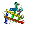
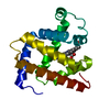
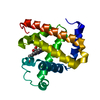
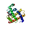
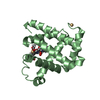
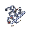
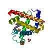
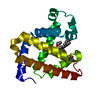
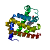
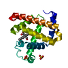
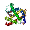
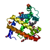
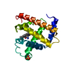
Entry Database : PDB / ID : 5jomTitle X-ray structure of CO-bound sperm whale myoglobin using a fixed target crystallography chip Myoglobin Keywords / / / Function / homology Function Domain/homology Component
/ / / / / / / / / / Biological species Physeter catodon (sperm whale)Method / / / Resolution : 1.9 Å Authors Oghbaey, S. / Sarracini, A. / Ginn, H.M. / Pare-Labrosse, O. / Kuo, A. / Marx, A. / Epp, S.W. / Sherrell, D.A. / Eger, B.T. / Zhong, Y. ...Oghbaey, S. / Sarracini, A. / Ginn, H.M. / Pare-Labrosse, O. / Kuo, A. / Marx, A. / Epp, S.W. / Sherrell, D.A. / Eger, B.T. / Zhong, Y. / Loch, R. / Mariani, V. / Alonso-Mori, R. / Nelson, S. / Lemke, H.T. / Owen, R.L. / Pearson, A.R. / Stuart, D.I. / Ernst, O.P. / Mueller-Werkmeister, H.M. / Miller, R.J.D. Funding support Organization Grant number Country Natural Sciences and Engineering Research Council (NSERC, Canada) Max Planck Society Canadian Institute for Advanced Research Canada Excellence Research Chair Program Anne and Max Tanenbaum Chair in Neuroscience at the University of Toronto People Programme (Marie Curie Actions) of the European Union's Seventh Framework Programme (FP7/2007-2013) REA grant agreement no. 623994 Department of Energy (DOE, United States) Contract No. DE-AC02-76SF00515
Journal : Acta Crystallogr D Struct Biol / Year : 2016Title : Fixed target combined with spectral mapping: approaching 100% hit rates for serial crystallography.Authors: Oghbaey, S. / Sarracini, A. / Ginn, H.M. / Pare-Labrosse, O. / Kuo, A. / Marx, A. / Epp, S.W. / Sherrell, D.A. / Eger, B.T. / Zhong, Y. / Loch, R. / Mariani, V. / Alonso-Mori, R. / Nelson, S. ... Authors : Oghbaey, S. / Sarracini, A. / Ginn, H.M. / Pare-Labrosse, O. / Kuo, A. / Marx, A. / Epp, S.W. / Sherrell, D.A. / Eger, B.T. / Zhong, Y. / Loch, R. / Mariani, V. / Alonso-Mori, R. / Nelson, S. / Lemke, H.T. / Owen, R.L. / Pearson, A.R. / Stuart, D.I. / Ernst, O.P. / Mueller-Werkmeister, H.M. / Miller, R.J. History Deposition May 2, 2016 Deposition site / Processing site Revision 1.0 Aug 17, 2016 Provider / Type Revision 1.1 Sep 13, 2017 Group / Derived calculations / Category / pdbx_struct_oper_listItem / _pdbx_struct_oper_list.symmetry_operationRevision 1.2 Nov 22, 2017 Group / Category / Item Revision 1.3 Feb 14, 2018 Group / Category Item / _diffrn_source.pdbx_synchrotron_siteRevision 1.4 Apr 18, 2018 Group / Category / Item Revision 1.5 Dec 4, 2019 Group / Category / Item Revision 1.6 Sep 27, 2023 Group / Database references / Refinement descriptionCategory chem_comp_atom / chem_comp_bond ... chem_comp_atom / chem_comp_bond / database_2 / diffrn_radiation_wavelength / pdbx_initial_refinement_model Item / _database_2.pdbx_database_accession
Show all Show less
 Movie
Movie Controller

 Yorodumi
 Open data
 Basic information
 Components
 Keywords
 Function and homology information
 X-RAY DIFFRACTION /
  Authors
 Canada,
  Germany,
  United States, 7items
  Citation
 Structure visualization
 Downloads & links
 Other downloads
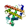


 PDBj
PDBj


 Assembly


 Mass: 616.487 Da / Num. of mol.: 1 / Source method: obtained synthetically / Formula: C34H32FeN4O4
 Mass: 96.063 Da / Num. of mol.: 1 / Source method: obtained synthetically / Formula: SO4
 Mass: 28.010 Da / Num. of mol.: 1 / Source method: obtained synthetically / Formula: CO
 Mass: 18.015 Da / Num. of mol.: 20 / Source method: isolated from a natural source / Formula: H2O
 Sample preparation
 Processing