 Movie
Movie Controller

[English] 日本語
 Yorodumi
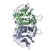
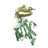
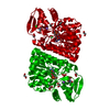
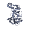
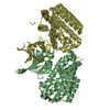
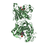
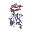
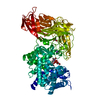
- PDB-5jmo: X-ray structure of furin in complex with the inhibitory antibody Nb14 -
+ Open data

- Basic information
| Entry | Database: PDB / ID: 5jmo | ||||||
|---|---|---|---|---|---|---|---|
| Title | X-ray structure of furin in complex with the inhibitory antibody Nb14 | ||||||
 Components Components |
| ||||||
 Keywords Keywords | Hydrolase/Antibody / protease / camelid / antibody / complex / Hydrolase-Antibody complex / furin | ||||||
| Function / homology |  Function and homology information Function and homology informationfurin / nerve growth factor production / dibasic protein processing / plasma lipoprotein particle remodeling / NGF processing / negative regulation of transforming growth factor beta1 production / Assembly of active LPL and LIPC lipase complexes / : / regulation of cholesterol transport / negative regulation of low-density lipoprotein particle receptor catabolic process ...furin / nerve growth factor production / dibasic protein processing / plasma lipoprotein particle remodeling / NGF processing / negative regulation of transforming growth factor beta1 production / Assembly of active LPL and LIPC lipase complexes / : / regulation of cholesterol transport / negative regulation of low-density lipoprotein particle receptor catabolic process / peptide biosynthetic process / Pre-NOTCH Processing in Golgi / nerve growth factor binding / Synthesis and processing of ENV and VPU / cytokine precursor processing / secretion by cell / Formation of the cornified envelope / Signaling by PDGF / trans-Golgi network transport vesicle / heparan sulfate binding / Signaling by NODAL / Elastic fibre formation / blastocyst formation / peptide hormone processing / positive regulation of membrane protein ectodomain proteolysis / CD163 mediating an anti-inflammatory response / zymogen activation / Regulation of CDH1 posttranslational processing and trafficking to plasma membrane / endopeptidase inhibitor activity / Activation of Matrix Metalloproteinases / Collagen degradation / collagen catabolic process / TGF-beta receptor signaling activates SMADs / regulation of protein catabolic process / Maturation of hRSV A proteins / Respiratory syncytial virus (RSV) attachment and entry / extracellular matrix disassembly / Uptake and function of anthrax toxins / regulation of signal transduction / Removal of aminoterminal propeptides from gamma-carboxylated proteins / endopeptidase activator activity / viral life cycle / peptide binding / extracellular matrix organization / transforming growth factor beta receptor signaling pathway / serine-type peptidase activity / cholesterol homeostasis / serine-type endopeptidase inhibitor activity / trans-Golgi network / negative regulation of inflammatory response to antigenic stimulus / protein maturation / SMAD2/SMAD3:SMAD4 heterotrimer regulates transcription / protein processing / Golgi lumen / peptidase activity / heparin binding / protease binding / endopeptidase activity / Induction of Cell-Cell Fusion / amyloid fibril formation / Potential therapeutics for SARS / positive regulation of viral entry into host cell / Attachment and Entry / endosome membrane / viral protein processing / membrane raft / Amyloid fiber formation / serine-type endopeptidase activity / Golgi membrane / cell surface / endoplasmic reticulum / extracellular exosome / extracellular region / membrane / metal ion binding / plasma membrane Similarity search - Function | ||||||
| Biological species |  Homo sapiens (human) Homo sapiens (human) synthetic construct (others) | ||||||
| Method |  X-RAY DIFFRACTION / SYNCHROTRON / MOLECULAR REPLACEMENT / Resolution: 1.998 Å X-RAY DIFFRACTION / SYNCHROTRON / MOLECULAR REPLACEMENT / Resolution: 1.998 Å | ||||||
 Authors Authors | Dahms, S.O. / Than, M.E. | ||||||
 Citation Citation | Journal: Sci Rep / Year: 2016 Title: The structure of a furin-antibody complex explains non-competitive inhibition by steric exclusion of substrate conformers. Authors: Dahms, S.O. / Creemers, J.W. / Schaub, Y. / Bourenkov, G.P. / Zogg, T. / Brandstetter, H. / Than, M.E. | ||||||
| History |
|
- Structure visualization
| Structure viewer | Molecule: MolmilJmol/JSmol |
|---|
- Downloads & links
-Download
| PDBx/mmCIF format | 5jmo.cif.gz | 260.5 KB | Display | PDBx/mmCIF format |
|---|---|---|---|---|
| PDB format | pdb5jmo.ent.gz | 203.4 KB | Display | PDB format |
| PDBx/mmJSON format | 5jmo.json.gz | Tree view | PDBx/mmJSON format | |
| Others |  Other downloads Other downloads |
-Validation report
| Arichive directory | https://data.pdbj.org/pub/pdb/validation_reports/jm/5jmoftp://data.pdbj.org/pub/pdb/validation_reports/jm/5jmo | HTTPS FTP |
|---|
-Related structure data
| Related structure data |  5jmrSC  4rydS S: Starting model for refinement C: citing same article ( |
|---|---|
| Similar structure data |


-Links
 PDBj
PDBj


- Assembly
| Deposited unit | 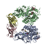
| |||||||||
|---|---|---|---|---|---|---|---|---|---|---|
| 1 | 
| |||||||||
| 2 | 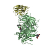
| |||||||||
| Unit cell |
| |||||||||
| Components on special symmetry positions |
|
-Components
-Protein / Antibody / Protein/peptide / Sugars , 4 types, 10 molecules ABCDGH
| #1: Protein | Mass: 52388.602 Da / Num. of mol.: 2 Source method: isolated from a genetically manipulated source Source: (gene. exp.) Homo sapiens (human) / Gene: FURIN, FUR, PACE, PCSK3 / Cell line (production host): HEK293S / Production host: Homo sapiens (human) / References: UniProt: P09958, furin#2: Antibody | Mass: 12615.001 Da / Num. of mol.: 2 Source method: isolated from a genetically manipulated source Source: (gene. exp.)  #3: Protein/peptide | 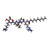  Type: Peptide-like / Class: Inhibitor / Mass: 749.452 Da / Num. of mol.: 2 / Source method: obtained synthetically / Source: (synth.) synthetic construct (others) / References: DECANOYL-ARG-VAL-LYS-ARG-CHLOROMETHYLKETONE Type: Peptide-like / Class: Inhibitor / Mass: 749.452 Da / Num. of mol.: 2 / Source method: obtained synthetically / Source: (synth.) synthetic construct (others) / References: DECANOYL-ARG-VAL-LYS-ARG-CHLOROMETHYLKETONE#4: Sugar | ChemComp-NAG /  Type: D-saccharide, beta linking / Mass: 221.208 Da / Num. of mol.: 4 Type: D-saccharide, beta linking / Mass: 221.208 Da / Num. of mol.: 4Source method: isolated from a genetically manipulated source Formula: C8H15NO6 |
|---|
-Non-polymers , 3 types, 777 molecules 


| #5: Chemical | ChemComp-CA /  Mass: 40.078 Da / Num. of mol.: 6 / Source method: obtained synthetically / Formula: Ca Mass: 40.078 Da / Num. of mol.: 6 / Source method: obtained synthetically / Formula: Ca#6: Chemical |  Mass: 22.990 Da / Num. of mol.: 2 / Source method: obtained synthetically / Formula: Na Mass: 22.990 Da / Num. of mol.: 2 / Source method: obtained synthetically / Formula: Na#7: Water | ChemComp-HOH / | Mass: 18.015 Da / Num. of mol.: 769 / Source method: isolated from a natural source / Formula: H2O |
|---|
-Experimental details
-Experiment
| Experiment | Method: X-RAY DIFFRACTION / Number of used crystals: 1 |
|---|
- Sample preparation
| Crystal | Density Matthews: 2.33 Å3/Da / Density % sol: 47.2 % |
|---|---|
| Crystal grow | Temperature: 293.15 K / Method: vapor diffusion, sitting drop / Details: .1 M sodium acetate, pH 5.6, 16-18% PEG 3350 |
-Data collection
| Diffraction | Mean temperature: 100 K |
|---|---|
| Diffraction source | Source: SYNCHROTRON / Site: PETRA III, EMBL c/o DESY  / Beamline: P13 (MX1) / Wavelength: 0.9763 Å / Beamline: P13 (MX1) / Wavelength: 0.9763 Å |
| Detector | Type: DECTRIS PILATUS 6M / Detector: PIXEL / Date: Jun 18, 2015 |
| Radiation | Protocol: SINGLE WAVELENGTH / Monochromatic (M) / Laue (L): M / Scattering type: x-ray |
| Radiation wavelength | Wavelength: 0.9763 Å / Relative weight: 1 |
| Reflection | Resolution: 1.998→66.381 Å / Num. obs: 83341 / % possible obs: 98.8 % / Redundancy: 6.73 % / Rsym value: 0.132 / Net I/σ(I): 12.02 |
| Reflection shell | Resolution: 1.998→2.12 Å / Rsym value: 0.608 |
- Processing
| Software |
| |||||||||||||||||||||||||||||||||||||||||||||||||||||||||||||||||||||||||||||||||||||||||||||||||||||||||||||||||||||||||||||||||||||||||||||||||||||||||||||||||||||||||||||||||||||||||||||||||||||||||||||||||||||||||
|---|---|---|---|---|---|---|---|---|---|---|---|---|---|---|---|---|---|---|---|---|---|---|---|---|---|---|---|---|---|---|---|---|---|---|---|---|---|---|---|---|---|---|---|---|---|---|---|---|---|---|---|---|---|---|---|---|---|---|---|---|---|---|---|---|---|---|---|---|---|---|---|---|---|---|---|---|---|---|---|---|---|---|---|---|---|---|---|---|---|---|---|---|---|---|---|---|---|---|---|---|---|---|---|---|---|---|---|---|---|---|---|---|---|---|---|---|---|---|---|---|---|---|---|---|---|---|---|---|---|---|---|---|---|---|---|---|---|---|---|---|---|---|---|---|---|---|---|---|---|---|---|---|---|---|---|---|---|---|---|---|---|---|---|---|---|---|---|---|---|---|---|---|---|---|---|---|---|---|---|---|---|---|---|---|---|---|---|---|---|---|---|---|---|---|---|---|---|---|---|---|---|---|---|---|---|---|---|---|---|---|---|---|---|---|---|---|---|---|
| Refinement | Method to determine structure: MOLECULAR REPLACEMENT Starting model: 4RYD (furin, chain A or chain B) and 5JMR (antibody, chain C or chain D) Resolution: 1.998→66.381 Å / SU ML: 0.17 / Cross valid method: FREE R-VALUE / σ(F): 1.21 / Phase error: 17.91 / Stereochemistry target values: ML
| |||||||||||||||||||||||||||||||||||||||||||||||||||||||||||||||||||||||||||||||||||||||||||||||||||||||||||||||||||||||||||||||||||||||||||||||||||||||||||||||||||||||||||||||||||||||||||||||||||||||||||||||||||||||||
| Solvent computation | Shrinkage radii: 0.9 Å / VDW probe radii: 1.11 Å / Solvent model: FLAT BULK SOLVENT MODEL | |||||||||||||||||||||||||||||||||||||||||||||||||||||||||||||||||||||||||||||||||||||||||||||||||||||||||||||||||||||||||||||||||||||||||||||||||||||||||||||||||||||||||||||||||||||||||||||||||||||||||||||||||||||||||
| Refinement step | Cycle: LAST / Resolution: 1.998→66.381 Å
| |||||||||||||||||||||||||||||||||||||||||||||||||||||||||||||||||||||||||||||||||||||||||||||||||||||||||||||||||||||||||||||||||||||||||||||||||||||||||||||||||||||||||||||||||||||||||||||||||||||||||||||||||||||||||
| Refine LS restraints |
| |||||||||||||||||||||||||||||||||||||||||||||||||||||||||||||||||||||||||||||||||||||||||||||||||||||||||||||||||||||||||||||||||||||||||||||||||||||||||||||||||||||||||||||||||||||||||||||||||||||||||||||||||||||||||
| LS refinement shell |
|
