 Movie
Movie Controller


+ Open data

- Basic information
| Entry | Database: PDB / ID: 5jk7 | ||||||
|---|---|---|---|---|---|---|---|
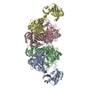
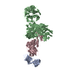
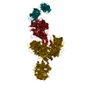
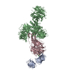
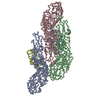
| Title | The X-ray structure of the DDB1-DCAF1-Vpr-UNG2 complex | ||||||
 Components Components |
| ||||||
 Keywords Keywords | Viral protein/DNA BINDING PROTEIN / Cullin4-RING E3 ubiquitin ligase HIV-1 Vpr UNG2 / DNA BINDING PROTEIN-HYDROLASE complex / Viral protein-DNA BINDING PROTEIN complex | ||||||
| Function / homology |  Function and homology information Function and homology informationcell competition in a multicellular organism / histone H2AT120 kinase activity / symbiont-mediated arrest of host cell cycle during G2/M transition / base-excision repair, AP site formation via deaminated base removal / uracil-DNA glycosylase / depyrimidination / Displacement of DNA glycosylase by APEX1 / single strand break repair / uracil DNA N-glycosylase activity / V(D)J recombination ...cell competition in a multicellular organism / histone H2AT120 kinase activity / symbiont-mediated arrest of host cell cycle during G2/M transition / base-excision repair, AP site formation via deaminated base removal / uracil-DNA glycosylase / depyrimidination / Displacement of DNA glycosylase by APEX1 / single strand break repair / uracil DNA N-glycosylase activity / V(D)J recombination / isotype switching / positive regulation by virus of viral protein levels in host cell / spindle assembly involved in female meiosis / epigenetic programming in the zygotic pronuclei / UV-damage excision repair / biological process involved in interaction with symbiont / regulation of mitotic cell cycle phase transition / WD40-repeat domain binding / Cul4A-RING E3 ubiquitin ligase complex / Cul4-RING E3 ubiquitin ligase complex / Cul4B-RING E3 ubiquitin ligase complex / ubiquitin ligase complex scaffold activity / negative regulation of reproductive process / negative regulation of developmental process / ribosomal small subunit binding / viral release from host cell / cullin family protein binding / ectopic germ cell programmed cell death / somatic hypermutation of immunoglobulin genes / positive regulation of viral genome replication / ubiquitin-like ligase-substrate adaptor activity / proteasomal protein catabolic process / post-translational protein modification / positive regulation of gluconeogenesis / Recognition and association of DNA glycosylase with site containing an affected pyrimidine / Cleavage of the damaged pyrimidine / B cell differentiation / nuclear estrogen receptor binding / Chromatin modifications during the maternal to zygotic transition (MZT) / symbiont-mediated activation of host apoptosis / nucleotide-excision repair / sperm end piece / protein homooligomerization / regulation of circadian rhythm / Recognition of DNA damage by PCNA-containing replication complex / base-excision repair / virion component / viral penetration into host nucleus / DNA Damage Recognition in GG-NER / Dual Incision in GG-NER / Transcription-Coupled Nucleotide Excision Repair (TC-NER) / fibrillar center / Wnt signaling pathway / Formation of TC-NER Pre-Incision Complex / Formation of Incision Complex in GG-NER / Dual incision in TC-NER / Gap-filling DNA repair synthesis and ligation in TC-NER / positive regulation of protein catabolic process / cellular response to UV / rhythmic process / host cell / Antigen processing: Ubiquitination & Proteasome degradation / site of double-strand break / sperm principal piece / Neddylation / sperm midpiece / host extracellular region / monoatomic ion transmembrane transport / ubiquitin-dependent protein catabolic process / damaged DNA binding / proteasome-mediated ubiquitin-dependent protein catabolic process / protein-macromolecule adaptor activity / chromosome, telomeric region / non-specific serine/threonine protein kinase / protein ubiquitination / protein serine kinase activity / DNA repair / apoptotic process / DNA damage response / symbiont entry into host cell / regulation of DNA-templated transcription / DNA-templated transcription / centrosome / negative regulation of apoptotic process / host cell nucleus / protein-containing complex binding / nucleolus / negative regulation of transcription by RNA polymerase II / protein-containing complex / mitochondrion / : / DNA binding / extracellular exosome / nucleoplasm / ATP binding / nucleus / cytoplasm Similarity search - Function | ||||||
| Biological species |  Homo sapiens (human) Homo sapiens (human) Human immunodeficiency virus type 1 group M subtype B Human immunodeficiency virus type 1 group M subtype B | ||||||
| Method |  X-RAY DIFFRACTION / SYNCHROTRON / Resolution: 3.49 Å X-RAY DIFFRACTION / SYNCHROTRON / Resolution: 3.49 Å | ||||||
 Authors Authors | Calero, G. / Ahn, J. / Wu, Y. | ||||||
| Funding support |  United States, 1items United States, 1items
| ||||||
 Citation Citation | Journal: Nat.Struct.Mol.Biol. / Year: 2016 Title: The DDB1-DCAF1-Vpr-UNG2 crystal structure reveals how HIV-1 Vpr steers human UNG2 toward destruction. Authors: Wu, Y. / Zhou, X. / Barnes, C.O. / DeLucia, M. / Cohen, A.E. / Gronenborn, A.M. / Ahn, J. / Calero, G. | ||||||
| History |
|
- Structure visualization
| Structure viewer | Molecule: MolmilJmol/JSmol |
|---|
- Downloads & links
-Download
| PDBx/mmCIF format | 5jk7.cif.gz | 1.4 MB | Display | PDBx/mmCIF format |
|---|---|---|---|---|
| PDB format | pdb5jk7.ent.gz | 1.2 MB | Display | PDB format |
| PDBx/mmJSON format | 5jk7.json.gz | Tree view | PDBx/mmJSON format | |
| Others |  Other downloads Other downloads |
-Validation report
| Arichive directory | https://data.pdbj.org/pub/pdb/validation_reports/jk/5jk7ftp://data.pdbj.org/pub/pdb/validation_reports/jk/5jk7 | HTTPS FTP |
|---|
-Related structure data
| Similar structure data |
|---|


-Links
 PDBj
PDBj


- Assembly
| Deposited unit | 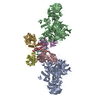
| ||||||||
|---|---|---|---|---|---|---|---|---|---|
| 1 | 
| ||||||||
| 2 | 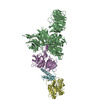
| ||||||||
| Unit cell |
|
-Components
| #1: Protein | Mass: 127097.469 Da / Num. of mol.: 2 Source method: isolated from a genetically manipulated source Source: (gene. exp.) Homo sapiens (human) / Gene: DDB1, XAP1Production host: Insect cell expression vector pTIE1 (others) References: UniProt: Q16531 #2: Protein | Mass: 41178.234 Da / Num. of mol.: 2 / Fragment: UNP residues 1045-1396 Source method: isolated from a genetically manipulated source Source: (gene. exp.) Homo sapiens (human) / Gene: VPRBP, DCAF1, KIAA0800, RIPProduction host: Insect cell expression vector pTIE1 (others) References: UniProt: Q9Y4B6, non-specific serine/threonine protein kinase #3: Protein | Mass: 25382.957 Da / Num. of mol.: 2 / Fragment: UNP residues 85-304 Source method: isolated from a genetically manipulated source Source: (gene. exp.) Homo sapiens (human) / Gene: UNG, DGU, UNG1, UNG15Production host: References: UniProt: P13051, uracil-DNA glycosylase #4: Protein | Mass: 11396.878 Da / Num. of mol.: 2 Source method: isolated from a genetically manipulated source Source: (gene. exp.) Human immunodeficiency virus type 1 group M subtype B (isolate NY5)Strain: isolate NY5 / Gene: vpr Production host: References: UniProt: P12520 |
|---|
-Experimental details
-Experiment
| Experiment | Method: X-RAY DIFFRACTION / Number of used crystals: 1 |
|---|
- Sample preparation
| Crystal | Density Matthews: 4.23 Å3/Da / Density % sol: 70.91 % |
|---|---|
| Crystal grow | Temperature: 289.15 K / Method: batch mode / Details: 100 mM Na Ctrate, pH 5.6, 11% PEG 20000 |
-Data collection
| Diffraction | Mean temperature: 100 K |
|---|---|
| Diffraction source | Source: SYNCHROTRON / Site: APS / Beamline: 23-ID-D / Wavelength: 0.979 Å |
| Detector | Type: MARMOSAIC 300 mm CCD / Detector: CCD / Date: Aug 14, 2015 |
| Radiation | Protocol: MAD / Monochromatic (M) / Laue (L): M / Scattering type: x-ray |
| Radiation wavelength | Wavelength: 0.979 Å / Relative weight: 1 |
| Reflection | Resolution: 3.49→40.3 Å / Num. obs: 77315 / Biso Wilson estimate: 188.78 Å2 |
- Processing
| Software |
| |||||||||||||||||||||||||||||||||||||||||||||||||||||||||||||||||||||||||||||||||||||||||||||||||||||||||||||||||||||||||||||||||||||||||||||||||||||||||||||||||||||||||||||||||||||||||||||||||||||||||||||||||||||||||||||||||
|---|---|---|---|---|---|---|---|---|---|---|---|---|---|---|---|---|---|---|---|---|---|---|---|---|---|---|---|---|---|---|---|---|---|---|---|---|---|---|---|---|---|---|---|---|---|---|---|---|---|---|---|---|---|---|---|---|---|---|---|---|---|---|---|---|---|---|---|---|---|---|---|---|---|---|---|---|---|---|---|---|---|---|---|---|---|---|---|---|---|---|---|---|---|---|---|---|---|---|---|---|---|---|---|---|---|---|---|---|---|---|---|---|---|---|---|---|---|---|---|---|---|---|---|---|---|---|---|---|---|---|---|---|---|---|---|---|---|---|---|---|---|---|---|---|---|---|---|---|---|---|---|---|---|---|---|---|---|---|---|---|---|---|---|---|---|---|---|---|---|---|---|---|---|---|---|---|---|---|---|---|---|---|---|---|---|---|---|---|---|---|---|---|---|---|---|---|---|---|---|---|---|---|---|---|---|---|---|---|---|---|---|---|---|---|---|---|---|---|---|---|---|---|---|---|---|---|
| Refinement | Resolution: 3.49→40.3 Å / Cor.coef. Fo:Fc: 0.9415 / Cor.coef. Fo:Fc free: 0.9297 / Cross valid method: THROUGHOUT / σ(F): 0 / SU Rfree Blow DPI: 0.422
| |||||||||||||||||||||||||||||||||||||||||||||||||||||||||||||||||||||||||||||||||||||||||||||||||||||||||||||||||||||||||||||||||||||||||||||||||||||||||||||||||||||||||||||||||||||||||||||||||||||||||||||||||||||||||||||||||
| Displacement parameters | Biso mean: 193.89 Å2
| |||||||||||||||||||||||||||||||||||||||||||||||||||||||||||||||||||||||||||||||||||||||||||||||||||||||||||||||||||||||||||||||||||||||||||||||||||||||||||||||||||||||||||||||||||||||||||||||||||||||||||||||||||||||||||||||||
| Refine analyze | Luzzati coordinate error obs: 1.036 Å | |||||||||||||||||||||||||||||||||||||||||||||||||||||||||||||||||||||||||||||||||||||||||||||||||||||||||||||||||||||||||||||||||||||||||||||||||||||||||||||||||||||||||||||||||||||||||||||||||||||||||||||||||||||||||||||||||
| Refinement step | Cycle: LAST / Resolution: 3.49→40.3 Å
| |||||||||||||||||||||||||||||||||||||||||||||||||||||||||||||||||||||||||||||||||||||||||||||||||||||||||||||||||||||||||||||||||||||||||||||||||||||||||||||||||||||||||||||||||||||||||||||||||||||||||||||||||||||||||||||||||
| Refine LS restraints |
| |||||||||||||||||||||||||||||||||||||||||||||||||||||||||||||||||||||||||||||||||||||||||||||||||||||||||||||||||||||||||||||||||||||||||||||||||||||||||||||||||||||||||||||||||||||||||||||||||||||||||||||||||||||||||||||||||
| LS refinement shell | Resolution: 3.49→3.58 Å / Total num. of bins used: 20
| |||||||||||||||||||||||||||||||||||||||||||||||||||||||||||||||||||||||||||||||||||||||||||||||||||||||||||||||||||||||||||||||||||||||||||||||||||||||||||||||||||||||||||||||||||||||||||||||||||||||||||||||||||||||||||||||||
| Refinement TLS params. | Method: refined / Refine-ID: X-RAY DIFFRACTION
| |||||||||||||||||||||||||||||||||||||||||||||||||||||||||||||||||||||||||||||||||||||||||||||||||||||||||||||||||||||||||||||||||||||||||||||||||||||||||||||||||||||||||||||||||||||||||||||||||||||||||||||||||||||||||||||||||
| Refinement TLS group |
|
