 Movie
Movie Controller


+ Open data

- Basic information
| Entry | Database: PDB / ID: 5jjg | ||||||
|---|---|---|---|---|---|---|---|
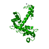
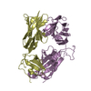
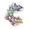
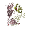
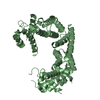
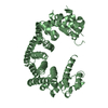
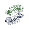
| Title | Structure of magnesium-loaded ALG-2 | ||||||
 Components Components | Pcalcium-binding protein ALG-2, rogrammed cell death protein 6 | ||||||
 Keywords Keywords |  APOPTOSIS / ALG-2 / penta-EF-HAND PROTEIN / CALCIUM BINDING PROTEIN / Apoptosis-Linked Gene 2 APOPTOSIS / ALG-2 / penta-EF-HAND PROTEIN / CALCIUM BINDING PROTEIN / Apoptosis-Linked Gene 2 | ||||||
| Function / homology |  Function and homology information Function and homology informationneural crest formation / vascular endothelial growth factor receptor-2 signaling pathway / neural crest cell development / COPII vesicle coat / COPII vesicle coating / negative regulation of TOR signaling / positive regulation of protein monoubiquitination / Cul3-RING ubiquitin ligase complex / negative regulation of vascular endothelial growth factor receptor signaling pathway / endoplasmic reticulum exit site ...neural crest formation / vascular endothelial growth factor receptor-2 signaling pathway / neural crest cell development / COPII vesicle coat / COPII vesicle coating / negative regulation of TOR signaling / positive regulation of protein monoubiquitination / Cul3-RING ubiquitin ligase complex / negative regulation of vascular endothelial growth factor receptor signaling pathway / endoplasmic reticulum exit site / ubiquitin-like ligase-substrate adaptor activity / negative regulation of phosphatidylinositol 3-kinase/protein kinase B signal transduction / endoplasmic reticulum to Golgi vesicle-mediated transport / protein-membrane adaptor activity / positive regulation of endothelial cell proliferation / positive regulation of endothelial cell migration / intracellular protein transport / response to calcium ion / positive regulation of angiogenesis / calcium-dependent protein binding / protein-macromolecule adaptor activity / cellular response to heat / cytoplasmic vesicle / angiogenesis / protein dimerization activity / endosome / apoptotic process / calcium ion binding / endoplasmic reticulum membrane / magnesium ion binding / endoplasmic reticulum / protein homodimerization activity / nucleoplasm / identical protein binding / nucleus / cytosol / cytoplasmSimilarity search - Function | ||||||
| Biological species |  Mus musculus (house mouse) Mus musculus (house mouse) | ||||||
| Method | X-RAY DIFFRACTION / SYNCHROTRON / MOLECULAR REPLACEMENT / Resolution: 1.72 Å | ||||||
 Authors Authors | Tanner, J.J. | ||||||
 Citation Citation | Journal: Biochemistry / Year: 2016 Title: EF5 Is the High-Affinity Mg(2+) Site in ALG-2. Authors: Tanner, J.J. / Frey, B.B. / Pemberton, T. / Henzl, M.T. | ||||||
| History |
|
- Structure visualization
| Structure viewer | Molecule: MolmilJmol/JSmol |
|---|
- Downloads & links
-Download
| PDBx/mmCIF format | 5jjg.cif.gz | 88.3 KB | Display | PDBx/mmCIF format |
|---|---|---|---|---|
| PDB format | pdb5jjg.ent.gz | 64.3 KB | Display | PDB format |
| PDBx/mmJSON format | 5jjg.json.gz | Tree view | PDBx/mmJSON format | |
| Others |  Other downloads Other downloads |
-Validation report
| Arichive directory | https://data.pdbj.org/pub/pdb/validation_reports/jj/5jjgftp://data.pdbj.org/pub/pdb/validation_reports/jj/5jjg | HTTPS FTP |
|---|
-Related structure data
| Related structure data | 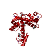 2zndS S: Starting model for refinement |
|---|---|
| Similar structure data |

-Links
 PDBj
PDBj
- Assembly
| Deposited unit | 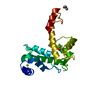
| ||||||||
|---|---|---|---|---|---|---|---|---|---|
| 1 | 
| ||||||||
| Unit cell |
|
-Components
| #1: Protein | Mass: 19950.262 Da / Num. of mol.: 1 Source method: isolated from a genetically manipulated source Source: (gene. exp.) Mus musculus (house mouse) / Gene: Pdcd6, Alg2 / Production host:  Escherichia coli (E. coli) / References: UniProt: P12815 Escherichia coli (E. coli) / References: UniProt: P12815 | ||||
|---|---|---|---|---|---|
| #2: Chemical |   Mass: 24.305 Da / Num. of mol.: 3 / Source method: obtained synthetically / Formula: Mg Mass: 24.305 Da / Num. of mol.: 3 / Source method: obtained synthetically / Formula: Mg#3: Chemical | Isopropyl alcohol  Mass: 60.095 Da / Num. of mol.: 3 / Source method: obtained synthetically / Formula: C3H8O / Comment: alkaloid*YM Mass: 60.095 Da / Num. of mol.: 3 / Source method: obtained synthetically / Formula: C3H8O / Comment: alkaloid*YM#4: Water | ChemComp-HOH / | Water Mass: 18.015 Da / Num. of mol.: 106 / Source method: isolated from a natural source / Formula: H2O Mass: 18.015 Da / Num. of mol.: 106 / Source method: isolated from a natural source / Formula: H2O |
-Experimental details
-Experiment
| Experiment | Method: X-RAY DIFFRACTION / Number of used crystals: 1 |
|---|
- Sample preparation
| Crystal | Density Matthews: 2.53 Å3/Da / Density % sol: 51.37 % |
|---|---|
| Crystal grow | Temperature: 277 K / Method: vapor diffusion, hanging drop / pH: 7.4 Details: 39-32% isopropanol, 0.10 M Tris, pH 7.4, 1.0 mM EGTA, 1.0 mM MgCl2 |
-Data collection
| Diffraction | Mean temperature: 100 K | |||||||||||||||
|---|---|---|---|---|---|---|---|---|---|---|---|---|---|---|---|---|
| Diffraction source | Source: SYNCHROTRON / Site: ALS  / Beamline: 4.2.2 / Wavelength: 1 Å / Beamline: 4.2.2 / Wavelength: 1 Å | |||||||||||||||
| Detector | Type: RDI CMOS_8M / Detector: CMOS / Date: Oct 10, 2015 | |||||||||||||||
| Radiation | Protocol: SINGLE WAVELENGTH / Monochromatic (M) / Laue (L): M / Scattering type: x-ray | |||||||||||||||
| Radiation wavelength | Wavelength: 1 Å / Relative weight: 1 | |||||||||||||||
| Reflection | Resolution: 1.72→54.35 Å / Num. obs: 22193 / % possible obs: 100 % / Redundancy: 6.9 % / Biso Wilson estimate: 24.81 Å2 / CC1/2: 1 / Rmerge(I) obs: 0.045 / Net I/σ(I): 25.8 | |||||||||||||||
| Reflection shell |
|
- Processing
| Software |
| ||||||||||||||||||||||||||||||||||||||||||||||||||||||||||||||||||||||||||||||||||||||||||||||||||||||||||||||||
|---|---|---|---|---|---|---|---|---|---|---|---|---|---|---|---|---|---|---|---|---|---|---|---|---|---|---|---|---|---|---|---|---|---|---|---|---|---|---|---|---|---|---|---|---|---|---|---|---|---|---|---|---|---|---|---|---|---|---|---|---|---|---|---|---|---|---|---|---|---|---|---|---|---|---|---|---|---|---|---|---|---|---|---|---|---|---|---|---|---|---|---|---|---|---|---|---|---|---|---|---|---|---|---|---|---|---|---|---|---|---|---|---|---|
| Refinement | Method to determine structure: MOLECULAR REPLACEMENT Starting model: 2ZND Resolution: 1.72→54.35 Å / SU ML: 0.2 / Cross valid method: FREE R-VALUE / σ(F): 1.26 / Phase error: 21.44
| ||||||||||||||||||||||||||||||||||||||||||||||||||||||||||||||||||||||||||||||||||||||||||||||||||||||||||||||||
| Solvent computation | Shrinkage radii: 0.9 Å / VDW probe radii: 1.11 Å | ||||||||||||||||||||||||||||||||||||||||||||||||||||||||||||||||||||||||||||||||||||||||||||||||||||||||||||||||
| Displacement parameters | Biso max: 86.59 Å2 / Biso mean: 30.2269 Å2 / Biso min: 14.78 Å2 | ||||||||||||||||||||||||||||||||||||||||||||||||||||||||||||||||||||||||||||||||||||||||||||||||||||||||||||||||
| Refinement step | Cycle: final / Resolution: 1.72→54.35 Å
| ||||||||||||||||||||||||||||||||||||||||||||||||||||||||||||||||||||||||||||||||||||||||||||||||||||||||||||||||
| Refine LS restraints |
| ||||||||||||||||||||||||||||||||||||||||||||||||||||||||||||||||||||||||||||||||||||||||||||||||||||||||||||||||
| LS refinement shell | Refine-ID: X-RAY DIFFRACTION / Total num. of bins used: 15
| ||||||||||||||||||||||||||||||||||||||||||||||||||||||||||||||||||||||||||||||||||||||||||||||||||||||||||||||||
| Refinement TLS params. | Method: refined / Origin x: -13.71 Å / Origin y: 16.8607 Å / Origin z: -18.6279 Å
| ||||||||||||||||||||||||||||||||||||||||||||||||||||||||||||||||||||||||||||||||||||||||||||||||||||||||||||||||
| Refinement TLS group | Selection details: chain A |
