 Movie
Movie Controller

[English] 日本語
 Yorodumi
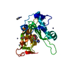
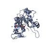
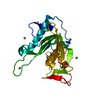
- PDB-5jf7: Crystal structure of type 2 PDF from Streptococcus agalactiae in ... -
+ Open data

- Basic information
| Entry | Database: PDB / ID: 5jf7 | ||||||
|---|---|---|---|---|---|---|---|
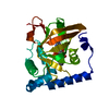
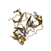
| Title | Crystal structure of type 2 PDF from Streptococcus agalactiae in complex with inhibitor SMP289 | ||||||
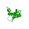
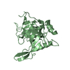
 Components Components | Peptide deformylase | ||||||
 Keywords Keywords | HYDROLASE / PDF / type 2 / NME / N-terminal methionine excision / Streptococcus agalactiae / inhibitor / SMP289 | ||||||
| Function / homology |  Function and homology information Function and homology informationpeptide deformylase / peptide deformylase activity / : / translation / metal ion binding Similarity search - Function | ||||||
| Biological species |  Streptococcus agalactiae (bacteria) Streptococcus agalactiae (bacteria) | ||||||
| Method |  X-RAY DIFFRACTION / SYNCHROTRON / MOLECULAR REPLACEMENT / Resolution: 2.1 Å X-RAY DIFFRACTION / SYNCHROTRON / MOLECULAR REPLACEMENT / Resolution: 2.1 Å | ||||||
 Authors Authors | Fieulaine, S. / Giglione, C. / Meinnel, T. / Hamiche, K. | ||||||
| Funding support |  France, 1items France, 1items
| ||||||
 Citation Citation | Journal: Sci Rep / Year: 2016 Title: A unique peptide deformylase platform to rationally design and challenge novel active compounds. Authors: Fieulaine, S. / Alves de Sousa, R. / Maigre, L. / Hamiche, K. / Alimi, M. / Bolla, J.M. / Taleb, A. / Denis, A. / Pages, J.M. / Artaud, I. / Meinnel, T. / Giglione, C. | ||||||
| History |
|
- Structure visualization
| Structure viewer | Molecule: MolmilJmol/JSmol |
|---|
- Downloads & links
-Download
| PDBx/mmCIF format | 5jf7.cif.gz | 100.2 KB | Display | PDBx/mmCIF format |
|---|---|---|---|---|
| PDB format | pdb5jf7.ent.gz | 75 KB | Display | PDB format |
| PDBx/mmJSON format | 5jf7.json.gz | Tree view | PDBx/mmJSON format | |
| Others |  Other downloads Other downloads |
-Validation report
| Summary document | 5jf7_validation.pdf.gz | 861.5 KB | Display | wwPDB validaton report |
|---|---|---|---|---|
| Full document | 5jf7_full_validation.pdf.gz | 863.2 KB | Display | |
| Data in XML | 5jf7_validation.xml.gz | 13.2 KB | Display | |
| Data in CIF | 5jf7_validation.cif.gz | 19.4 KB | Display | |
| Arichive directory | https://data.pdbj.org/pub/pdb/validation_reports/jf/5jf7ftp://data.pdbj.org/pub/pdb/validation_reports/jf/5jf7 | HTTPS FTP |
-Related structure data
| Related structure data |  5jexC  5jeyC  5jezC  5jf0C  5jf1C  5jf2C  5jf3C  5jf4C  5jf5C  5jf6C  5jf8C 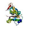 2aieS S: Starting model for refinement C: citing same article ( |
|---|---|
| Similar structure data |


-Links
 PDBj
PDBj


- Assembly
| Deposited unit | 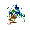
| ||||||||
|---|---|---|---|---|---|---|---|---|---|
| 1 |
| ||||||||
| Unit cell |
|
-Components
-Protein , 1 types, 1 molecules A
| #1: Protein | Mass: 22894.297 Da / Num. of mol.: 1 / Mutation: S2A Source method: isolated from a genetically manipulated source Source: (gene. exp.) Streptococcus agalactiae (bacteria) / Gene: def, gbs1883 / Production host: |
|---|
-Non-polymers , 5 types, 245 molecules 


| #2: Chemical | ChemComp-6JU /  Mass: 359.217 Da / Num. of mol.: 1 / Source method: obtained synthetically / Formula: C17H15BrN2O2 Mass: 359.217 Da / Num. of mol.: 1 / Source method: obtained synthetically / Formula: C17H15BrN2O2 | ||
|---|---|---|---|
| #3: Chemical | ChemComp-ACT /  Mass: 59.044 Da / Num. of mol.: 1 / Source method: obtained synthetically / Formula: C2H3O2 Mass: 59.044 Da / Num. of mol.: 1 / Source method: obtained synthetically / Formula: C2H3O2 | ||
| #4: Chemical | ChemComp-IMD /  Mass: 69.085 Da / Num. of mol.: 1 / Source method: obtained synthetically / Formula: C3H5N2 Mass: 69.085 Da / Num. of mol.: 1 / Source method: obtained synthetically / Formula: C3H5N2 | ||
| #5: Chemical | ChemComp-ZN /  Mass: 65.409 Da / Num. of mol.: 9 / Source method: obtained synthetically / Formula: Zn Mass: 65.409 Da / Num. of mol.: 9 / Source method: obtained synthetically / Formula: Zn#6: Water | ChemComp-HOH / | Mass: 18.015 Da / Num. of mol.: 233 / Source method: isolated from a natural source / Formula: H2O |
-Experimental details
-Experiment
| Experiment | Method: X-RAY DIFFRACTION / Number of used crystals: 1 |
|---|
- Sample preparation
| Crystal | Density Matthews: 2.67 Å3/Da / Density % sol: 53.9 % |
|---|---|
| Crystal grow | Temperature: 293 K / Method: vapor diffusion, hanging drop / pH: 6.5 Details: 13% PEG-8000, 100mM Imidazole pH6.5, 400mM Zn acetate |
-Data collection
| Diffraction | Mean temperature: 100 K |
|---|---|
| Diffraction source | Source: SYNCHROTRON / Site: ESRF / Beamline: ID23-2 / Wavelength: 0.873 Å |
| Detector | Type: MARMOSAIC 225 mm CCD / Detector: CCD / Date: Jan 28, 2010 |
| Radiation | Protocol: SINGLE WAVELENGTH / Monochromatic (M) / Laue (L): M / Scattering type: x-ray |
| Radiation wavelength | Wavelength: 0.873 Å / Relative weight: 1 |
| Reflection | Resolution: 2.09→50 Å / Num. obs: 26386 / % possible obs: 94.8 % / Observed criterion σ(F): 2 / Observed criterion σ(I): 2 / Redundancy: 3.2 % / Rsym value: 0.093 / Net I/σ(I): 12.04 |
| Reflection shell | Resolution: 2.09→2.22 Å / Redundancy: 3.16 % / Rmerge(I) obs: 0.322 / Mean I/σ(I) obs: 3.99 / Rsym value: 0.381 / % possible all: 86.3 |
- Processing
| Software |
| ||||||||||||||||||||||||||||||||||||||||||||||||||||||||||||||||||||||||||||||||||||||||||||||||||||||||||||||||||||||||||||||||||||||||||||||||||||||||||||||||||||||||||||||||||||||
|---|---|---|---|---|---|---|---|---|---|---|---|---|---|---|---|---|---|---|---|---|---|---|---|---|---|---|---|---|---|---|---|---|---|---|---|---|---|---|---|---|---|---|---|---|---|---|---|---|---|---|---|---|---|---|---|---|---|---|---|---|---|---|---|---|---|---|---|---|---|---|---|---|---|---|---|---|---|---|---|---|---|---|---|---|---|---|---|---|---|---|---|---|---|---|---|---|---|---|---|---|---|---|---|---|---|---|---|---|---|---|---|---|---|---|---|---|---|---|---|---|---|---|---|---|---|---|---|---|---|---|---|---|---|---|---|---|---|---|---|---|---|---|---|---|---|---|---|---|---|---|---|---|---|---|---|---|---|---|---|---|---|---|---|---|---|---|---|---|---|---|---|---|---|---|---|---|---|---|---|---|---|---|---|
| Refinement | Method to determine structure: MOLECULAR REPLACEMENT Starting model: 2AIE Resolution: 2.1→44.66 Å / Cor.coef. Fo:Fc: 0.962 / Cor.coef. Fo:Fc free: 0.93 / SU B: 7.174 / SU ML: 0.111 / Cross valid method: THROUGHOUT / ESU R: 0.172 / ESU R Free: 0.163 / Stereochemistry target values: MAXIMUM LIKELIHOOD / Details: HYDROGENS HAVE BEEN ADDED IN THE RIDING POSITIONS
| ||||||||||||||||||||||||||||||||||||||||||||||||||||||||||||||||||||||||||||||||||||||||||||||||||||||||||||||||||||||||||||||||||||||||||||||||||||||||||||||||||||||||||||||||||||||
| Solvent computation | Ion probe radii: 0.8 Å / Shrinkage radii: 0.8 Å / VDW probe radii: 1.2 Å / Solvent model: MASK | ||||||||||||||||||||||||||||||||||||||||||||||||||||||||||||||||||||||||||||||||||||||||||||||||||||||||||||||||||||||||||||||||||||||||||||||||||||||||||||||||||||||||||||||||||||||
| Displacement parameters | Biso mean: 22.14 Å2
| ||||||||||||||||||||||||||||||||||||||||||||||||||||||||||||||||||||||||||||||||||||||||||||||||||||||||||||||||||||||||||||||||||||||||||||||||||||||||||||||||||||||||||||||||||||||
| Refinement step | Cycle: LAST / Resolution: 2.1→44.66 Å
| ||||||||||||||||||||||||||||||||||||||||||||||||||||||||||||||||||||||||||||||||||||||||||||||||||||||||||||||||||||||||||||||||||||||||||||||||||||||||||||||||||||||||||||||||||||||
| Refine LS restraints |
|