 Movie
Movie Controller

[English] 日本語
 Yorodumi
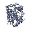
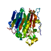
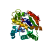
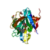
- PDB-5jd6: Crystal structure of MGS-MChE2, an alpha/beta hydrolase enzyme fr... -
+ Open data

- Basic information
| Entry | Database: PDB / ID: 5jd6 | ||||||
|---|---|---|---|---|---|---|---|
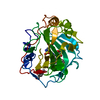
| Title | Crystal structure of MGS-MChE2, an alpha/beta hydrolase enzyme from the metagenome of sediments from the lagoon of Mar Chica, Morocco | ||||||
 Components Components | MGS-MChE2 | ||||||
 Keywords Keywords | HYDROLASE / alpha/beta hydrolase / esterase / metagenome | ||||||
| Function / homology | Alpha/Beta hydrolase fold, catalytic domain / Rossmann fold / 3-Layer(aba) Sandwich / Alpha Beta Function and homology information Function and homology information | ||||||
| Biological species |  uncultured bacterium (environmental samples) uncultured bacterium (environmental samples) | ||||||
| Method |  X-RAY DIFFRACTION / SYNCHROTRON / MOLECULAR REPLACEMENT / Resolution: 2.463 Å X-RAY DIFFRACTION / SYNCHROTRON / MOLECULAR REPLACEMENT / Resolution: 2.463 Å | ||||||
 Authors Authors | Stogios, P.J. / Xu, X. / Nocek, B. / Yim, V. / Cui, H. / Martinez-Martinez, M. / Golyshin, P.N. / Yakima, M.M. / Ferrer, M. / Savchenko, A. | ||||||
 Citation Citation | Journal: To Be Published Title: Crystal structure of MGS-MChE2, an alpha/beta hydrolase enzyme from the metagenome of sediments from the lagoon of Mar Chica, Morocco Authors: Martinez-Martinez, M. | ||||||
| History |
|
- Structure visualization
| Structure viewer | Molecule: MolmilJmol/JSmol |
|---|
- Downloads & links
-Download
| PDBx/mmCIF format | 5jd6.cif.gz | 102.7 KB | Display | PDBx/mmCIF format |
|---|---|---|---|---|
| PDB format | pdb5jd6.ent.gz | 78.4 KB | Display | PDB format |
| PDBx/mmJSON format | 5jd6.json.gz | Tree view | PDBx/mmJSON format | |
| Others |  Other downloads Other downloads |
-Validation report
| Arichive directory | https://data.pdbj.org/pub/pdb/validation_reports/jd/5jd6ftp://data.pdbj.org/pub/pdb/validation_reports/jd/5jd6 | HTTPS FTP |
|---|
-Related structure data
| Related structure data | 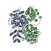 1cr6S S: Starting model for refinement |
|---|---|
| Similar structure data |


-Links
 PDBj
PDBj- Assembly
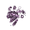
| Deposited unit | 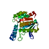
| ||||||||
|---|---|---|---|---|---|---|---|---|---|
| 1 |
| ||||||||
| Unit cell |
|
-Components
| #1: Protein | Mass: 28903.330 Da / Num. of mol.: 1 Source method: isolated from a genetically manipulated source Source: (gene. exp.) uncultured bacterium (environmental samples)Plasmid: p15Tv lic / Production host: |
|---|---|
| #2: Water | ChemComp-HOH /  Mass: 18.015 Da / Num. of mol.: 23 / Source method: isolated from a natural source / Formula: H2O Mass: 18.015 Da / Num. of mol.: 23 / Source method: isolated from a natural source / Formula: H2O |
-Experimental details
-Experiment
| Experiment | Method: X-RAY DIFFRACTION / Number of used crystals: 1 |
|---|
- Sample preparation
| Crystal | Density Matthews: 2.44 Å3/Da / Density % sol: 49.61 % |
|---|---|
| Crystal grow | Temperature: 298 K / Method: vapor diffusion, sitting drop / pH: 7 Details: 2 M ammonium sulphate, 5% isopropanol. Cryoprotectant: paratone-N oil. |
-Data collection
| Diffraction | Mean temperature: 100 K |
|---|---|
| Diffraction source | Source: SYNCHROTRON / Site: APS  / Beamline: 19-ID / Wavelength: 0.97915 Å / Beamline: 19-ID / Wavelength: 0.97915 Å |
| Detector | Type: ADSC QUANTUM 315r / Detector: CCD / Date: Feb 16, 2014 |
| Radiation | Protocol: SINGLE WAVELENGTH / Monochromatic (M) / Laue (L): M / Scattering type: x-ray |
| Radiation wavelength | Wavelength: 0.97915 Å / Relative weight: 1 |
| Reflection | Resolution: 2.47→25.848 Å / Num. obs: 10194 / % possible obs: 96.5 % / Redundancy: 6.8 % / Rmerge(I) obs: 0.036 / Net I/σ(I): 35.92 |
| Reflection shell | Resolution: 2.47→2.51 Å / Redundancy: 6.8 % / Rmerge(I) obs: 0.578 / % possible all: 99.2 |
- Processing
| Software |
| ||||||||||||||||||||||||||||||||||||||||||||||||||||||||||||||||||||||||||||||||||||||||||||||||||||||||||||||||||||||||||||||||||||||||||||||||||||||
|---|---|---|---|---|---|---|---|---|---|---|---|---|---|---|---|---|---|---|---|---|---|---|---|---|---|---|---|---|---|---|---|---|---|---|---|---|---|---|---|---|---|---|---|---|---|---|---|---|---|---|---|---|---|---|---|---|---|---|---|---|---|---|---|---|---|---|---|---|---|---|---|---|---|---|---|---|---|---|---|---|---|---|---|---|---|---|---|---|---|---|---|---|---|---|---|---|---|---|---|---|---|---|---|---|---|---|---|---|---|---|---|---|---|---|---|---|---|---|---|---|---|---|---|---|---|---|---|---|---|---|---|---|---|---|---|---|---|---|---|---|---|---|---|---|---|---|---|---|---|---|---|
| Refinement | Method to determine structure: MOLECULAR REPLACEMENT Starting model: 1CR6 Resolution: 2.463→25.848 Å / SU ML: 0.25 / Cross valid method: FREE R-VALUE / σ(F): 1.34 / Phase error: 26.1 / Stereochemistry target values: ML
| ||||||||||||||||||||||||||||||||||||||||||||||||||||||||||||||||||||||||||||||||||||||||||||||||||||||||||||||||||||||||||||||||||||||||||||||||||||||
| Solvent computation | Shrinkage radii: 0.9 Å / VDW probe radii: 1.11 Å / Solvent model: FLAT BULK SOLVENT MODEL | ||||||||||||||||||||||||||||||||||||||||||||||||||||||||||||||||||||||||||||||||||||||||||||||||||||||||||||||||||||||||||||||||||||||||||||||||||||||
| Refinement step | Cycle: LAST / Resolution: 2.463→25.848 Å
| ||||||||||||||||||||||||||||||||||||||||||||||||||||||||||||||||||||||||||||||||||||||||||||||||||||||||||||||||||||||||||||||||||||||||||||||||||||||
| Refine LS restraints |
| ||||||||||||||||||||||||||||||||||||||||||||||||||||||||||||||||||||||||||||||||||||||||||||||||||||||||||||||||||||||||||||||||||||||||||||||||||||||
| LS refinement shell |
| ||||||||||||||||||||||||||||||||||||||||||||||||||||||||||||||||||||||||||||||||||||||||||||||||||||||||||||||||||||||||||||||||||||||||||||||||||||||
| Refinement TLS params. | Method: refined / Refine-ID: X-RAY DIFFRACTION
| ||||||||||||||||||||||||||||||||||||||||||||||||||||||||||||||||||||||||||||||||||||||||||||||||||||||||||||||||||||||||||||||||||||||||||||||||||||||
| Refinement TLS group |
|
