 Movie
Movie Controller

[English] 日本語
 Yorodumi
- PDB-1cr6: CRYSTAL STRUCTURE OF MURINE SOLUBLE EPOXIDE HYDROLASE COMPLEXED W... -
+ Open data

- Basic information
| Entry | Database: PDB / ID: 1cr6 | ||||||
|---|---|---|---|---|---|---|---|
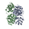
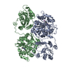
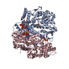
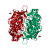
| Title | CRYSTAL STRUCTURE OF MURINE SOLUBLE EPOXIDE HYDROLASE COMPLEXED WITH CPU INHIBITOR | ||||||
 Components Components | EPOXIDE HYDROLASE | ||||||
 Keywords Keywords | HYDROLASE / HOMODIMER / ALPHA/BETA HYDROLASE FOLD / DISUBSTITUTED UREA INHIBITOR | ||||||
| Function / homology |  Function and homology information Function and homology informationSynthesis of epoxy (EET) and dihydroxyeicosatrienoic acids (DHET) / Biosynthesis of maresins / prostaglandin production involved in inflammatory response / lipid-phosphate phosphatase / 10-hydroxy-9-(phosphonooxy)octadecanoate phosphatase activity / stilbene catabolic process / epoxide metabolic process / phospholipid dephosphorylation / lipid phosphatase activity / soluble epoxide hydrolase ...Synthesis of epoxy (EET) and dihydroxyeicosatrienoic acids (DHET) / Biosynthesis of maresins / prostaglandin production involved in inflammatory response / lipid-phosphate phosphatase / 10-hydroxy-9-(phosphonooxy)octadecanoate phosphatase activity / stilbene catabolic process / epoxide metabolic process / phospholipid dephosphorylation / lipid phosphatase activity / soluble epoxide hydrolase / Peroxisomal protein import / lysophosphatidic acid phosphatase activity / linoleic acid metabolic process / positive regulation of blood pressure / epoxide hydrolase activity / dephosphorylation / regulation of cholesterol metabolic process / phosphatase activity / toxic substance binding / cholesterol homeostasis / response to toxic substance / peroxisome / positive regulation of gene expression / magnesium ion binding / protein homodimerization activity / cytosol Similarity search - Function | ||||||
| Biological species |  | ||||||
| Method |  X-RAY DIFFRACTION / SYNCHROTRON / Resolution: 2.8 Å X-RAY DIFFRACTION / SYNCHROTRON / Resolution: 2.8 Å | ||||||
 Authors Authors | Argiriadi, M.A. / Morisseau, C. / Hammock, B.D. / Christianson, D.W. | ||||||
 Citation Citation | Journal: Proc.Natl.Acad.Sci.USA / Year: 1999 Title: Detoxification of environmental mutagens and carcinogens: structure, mechanism, and evolution of liver epoxide hydrolase. Authors: Argiriadi, M.A. / Morisseau, C. / Hammock, B.D. / Christianson, D.W. #1: Journal: Proc.Natl.Acad.Sci.USA / Year: 1999Title: Potent urea and carbamate inhibitors of soluble epoxide hydrolases Authors: Morisseau, C. / Goodrow, M. / Dowdy, D. / Zheng, J. / Greene, J. / Sanborn, J.R. / Hammock, B.D. | ||||||
| History |
|
- Structure visualization
| Structure viewer | Molecule: MolmilJmol/JSmol |
|---|
- Downloads & links
-Download
| PDBx/mmCIF format | 1cr6.cif.gz | 212.7 KB | Display | PDBx/mmCIF format |
|---|---|---|---|---|
| PDB format | pdb1cr6.ent.gz | 170.4 KB | Display | PDB format |
| PDBx/mmJSON format | 1cr6.json.gz | Tree view | PDBx/mmJSON format | |
| Others |  Other downloads Other downloads |
-Validation report
| Arichive directory | https://data.pdbj.org/pub/pdb/validation_reports/cr/1cr6ftp://data.pdbj.org/pub/pdb/validation_reports/cr/1cr6 | HTTPS FTP |
|---|
-Related structure data


-Links
 PDBj
PDBj


- Assembly
| Deposited unit | 
| ||||||||
|---|---|---|---|---|---|---|---|---|---|
| 1 |
| ||||||||
| Unit cell |
| ||||||||
| Details | homodimer, each monomer represented by chain A and chain B related by an NCS two-fold. / domain-swapped dimer |
-Components
| #1: Protein | Mass: 62582.039 Da / Num. of mol.: 2 Source method: isolated from a genetically manipulated source Source: (gene. exp.)   Spodoptera frugiperda (fall armyworm) / References: UniProt: P34914, epoxide hydrolase Spodoptera frugiperda (fall armyworm) / References: UniProt: P34914, epoxide hydrolase#2: Chemical |   Mass: 260.375 Da / Num. of mol.: 2 / Source method: obtained synthetically / Formula: C16H24N2O Mass: 260.375 Da / Num. of mol.: 2 / Source method: obtained synthetically / Formula: C16H24N2O#3: Water | ChemComp-HOH / |  Mass: 18.015 Da / Num. of mol.: 21 / Source method: isolated from a natural source / Formula: H2O Mass: 18.015 Da / Num. of mol.: 21 / Source method: isolated from a natural source / Formula: H2O |
|---|
-Experimental details
-Experiment
| Experiment | Method: X-RAY DIFFRACTION / Number of used crystals: 1 |
|---|
- Sample preparation
| Crystal | Density Matthews: 2.6 Å3/Da / Density % sol: 52.72 % | |||||||||||||||||||||||||||||||||||||||||||||
|---|---|---|---|---|---|---|---|---|---|---|---|---|---|---|---|---|---|---|---|---|---|---|---|---|---|---|---|---|---|---|---|---|---|---|---|---|---|---|---|---|---|---|---|---|---|---|
| Crystal grow | Temperature: 298 K / Method: vapor diffusion / pH: 6 Details: ammonium sulfate, MES, ethanol, dithiothreitol, CPU (N-cyclohexyl-N'-(3-phenyl)propyl urea), pH 6.0, VAPOR DIFFUSION, temperature 298.0K | |||||||||||||||||||||||||||||||||||||||||||||
| Crystal grow | *PLUS Method: vapor diffusion, hanging drop | |||||||||||||||||||||||||||||||||||||||||||||
| Components of the solutions | *PLUS
|
-Data collection
| Diffraction | Mean temperature: 100 K |
|---|---|
| Diffraction source | Source: SYNCHROTRON / Site: CHESS  / Beamline: A1 / Wavelength: 0.893 / Beamline: A1 / Wavelength: 0.893 |
| Detector | Type: OTHER / Detector: CCD / Date: Aug 30, 1998 |
| Radiation | Protocol: SINGLE WAVELENGTH / Monochromatic (M) / Laue (L): M / Scattering type: x-ray |
| Radiation wavelength | Wavelength: 0.893 Å / Relative weight: 1 |
| Reflection | Resolution: 2.8→20 Å / Num. all: 73807 / Num. obs: 24877 / % possible obs: 0.916 % / Observed criterion σ(F): 1 / Observed criterion σ(I): 1 / Redundancy: 2.9 % / Biso Wilson estimate: 44.8 Å2 / Rmerge(I) obs: 0.096 / Net I/σ(I): 9.9 |
| Reflection shell | Resolution: 2.8→20 Å / Redundancy: 1 % / Rmerge(I) obs: 0.345 / Num. unique all: 1300 / % possible all: 0.428 |
| Reflection | *PLUS % possible obs: 80.1 % / Num. measured all: 73807 |
| Reflection shell | *PLUS % possible obs: 42.8 % |
- Processing
| Software |
| |||||||||||||||||||||||||
|---|---|---|---|---|---|---|---|---|---|---|---|---|---|---|---|---|---|---|---|---|---|---|---|---|---|---|
| Refinement | Resolution: 2.8→20 Å / σ(F): 2 / Stereochemistry target values: Engh & Huber
| |||||||||||||||||||||||||
| Refinement step | Cycle: LAST / Resolution: 2.8→20 Å
| |||||||||||||||||||||||||
| Refine LS restraints |
| |||||||||||||||||||||||||
| Software | *PLUS Name: X-PLOR / Version: 3.843 / Classification: refinement | |||||||||||||||||||||||||
| Refinement | *PLUS Highest resolution: 2.8 Å / σ(F): 2 / % reflection Rfree: 5 % / Rfactor obs: 0.201 | |||||||||||||||||||||||||
| Solvent computation | *PLUS | |||||||||||||||||||||||||
| Displacement parameters | *PLUS | |||||||||||||||||||||||||
| Refine LS restraints | *PLUS
|
