 Movie
Movie Controller


+ Open data

- Basic information
| Entry | Database: PDB / ID: 5jcg | ||||||
|---|---|---|---|---|---|---|---|
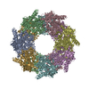
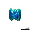
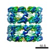
| Title | Structure of Human Peroxiredoxin 3 as three stacked rings | ||||||
 Components Components | Thioredoxin-dependent peroxide reductase, mitochondrial | ||||||
 Keywords Keywords | OXIDOREDUCTASE / Peroxidase / molecular chaperone / peroxiredoxin | ||||||
| Function / homology |  Function and homology information Function and homology informationnegative regulation of kinase activity / NADH-dependent peroxiredoxin activity / maternal placenta development / thioredoxin-dependent peroxiredoxin / thioredoxin peroxidase activity / myeloid cell differentiation / cysteine-type endopeptidase inhibitor activity involved in apoptotic process / Detoxification of Reactive Oxygen Species / cell redox homeostasis / : ...negative regulation of kinase activity / NADH-dependent peroxiredoxin activity / maternal placenta development / thioredoxin-dependent peroxiredoxin / thioredoxin peroxidase activity / myeloid cell differentiation / cysteine-type endopeptidase inhibitor activity involved in apoptotic process / Detoxification of Reactive Oxygen Species / cell redox homeostasis / : / hydrogen peroxide catabolic process / regulation of mitochondrial membrane potential / response to hydrogen peroxide / cellular response to reactive oxygen species / mitochondrion organization / cellular response to oxidative stress / response to oxidative stress / response to lipopolysaccharide / early endosome / mitochondrial matrix / positive regulation of cell population proliferation / protein kinase binding / negative regulation of apoptotic process / protein-containing complex / mitochondrion / nucleoplasm / identical protein binding / plasma membrane / cytoplasm / cytosol Similarity search - Function | ||||||
| Biological species |  Homo sapiens (human) Homo sapiens (human) | ||||||
| Method |  X-RAY DIFFRACTION / SYNCHROTRON / MOLECULAR REPLACEMENT / Resolution: 2.8 Å X-RAY DIFFRACTION / SYNCHROTRON / MOLECULAR REPLACEMENT / Resolution: 2.8 Å | ||||||
 Authors Authors | Yewdall, N.A. / Gerrard, J.A. / Goldstone, D.C. | ||||||
| Funding support |  New Zealand, 1items New Zealand, 1items
| ||||||
 Citation Citation | Journal: Structure / Year: 2016 Title: Structures of Human Peroxiredoxin 3 Suggest Self-Chaperoning Assembly that Maintains Catalytic State. Authors: N Amy Yewdall / Hariprasad Venugopal / Ambroise Desfosses / Vahid Abrishami / Yuliana Yosaatmadja / Mark B Hampton / Juliet A Gerrard / David C Goldstone / Alok K Mitra / Mazdak Radjainia /  Abstract: Peroxiredoxins are antioxidant proteins primarily responsible for detoxification of hydroperoxides in cells. On exposure to various cellular stresses, peroxiredoxins can acquire chaperone activity, ...Peroxiredoxins are antioxidant proteins primarily responsible for detoxification of hydroperoxides in cells. On exposure to various cellular stresses, peroxiredoxins can acquire chaperone activity, manifested as quaternary reorganization into a high molecular weight (HMW) form. Acidification, for example, causes dodecameric rings of human peroxiredoxin 3 (HsPrx3) to stack into long helical filaments. In this work, a 4.1-Å resolution structure of low-pH-instigated helical filaments was elucidated, showing a locally unfolded active site and partially folded C terminus. A 2.8-Å crystal structure of HsPrx3 was determined at pH 8.5 under reducing conditions, wherein dodecameric rings are arranged as a short stack, with symmetry similar to low-pH filaments. In contrast to previous observations, the crystal structure displays both a fully folded active site and ordered C terminus, suggesting that the HsPrx3 HMW form maintains catalytic activity. We propose a new role for the HMW form as a self-chaperoning assembly maintaining HsPrx3 function under stress. | ||||||
| History |
|
- Structure visualization
| Structure viewer | Molecule: MolmilJmol/JSmol |
|---|
- Downloads & links
-Download
| PDBx/mmCIF format | 5jcg.cif.gz | 337.4 KB | Display | PDBx/mmCIF format |
|---|---|---|---|---|
| PDB format | pdb5jcg.ent.gz | 274.8 KB | Display | PDB format |
| PDBx/mmJSON format | 5jcg.json.gz | Tree view | PDBx/mmJSON format | |
| Others |  Other downloads Other downloads |
-Validation report
| Arichive directory | https://data.pdbj.org/pub/pdb/validation_reports/jc/5jcgftp://data.pdbj.org/pub/pdb/validation_reports/jc/5jcg | HTTPS FTP |
|---|
-Related structure data
| Related structure data | 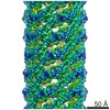 3414C  1zyeS S: Starting model for refinement C: citing same article ( |
|---|---|
| Similar structure data |


-Links
 PDBj
PDBj


- Assembly
| Deposited unit | 
| ||||||||
|---|---|---|---|---|---|---|---|---|---|
| 1 | 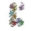
| ||||||||
| Unit cell |
|
-Components
| #1: Protein | Mass: 22194.162 Da / Num. of mol.: 9 Source method: isolated from a genetically manipulated source Source: (gene. exp.) Homo sapiens (human) / Gene: PRDX3, AOP1 / Plasmid: pET151-D-TOPO / Production host:  #2: Water | ChemComp-HOH / |  Mass: 18.015 Da / Num. of mol.: 159 / Source method: isolated from a natural source / Formula: H2O Mass: 18.015 Da / Num. of mol.: 159 / Source method: isolated from a natural source / Formula: H2O |
|---|
-Experimental details
-Experiment
| Experiment | Method: X-RAY DIFFRACTION / Number of used crystals: 1 |
|---|
- Sample preparation
| Crystal | Density Matthews: 3.21 Å3/Da / Density % sol: 61.69 % |
|---|---|
| Crystal grow | Temperature: 293 K / Method: vapor diffusion, sitting drop / pH: 8.5 Details: 12.5% PEG1000, 12.5% PEG3350. 12.5% MPD, 0.02 M alcohol additives at pH 8.5 (Gorrec, 2009) |
-Data collection
| Diffraction | Mean temperature: 100 K |
|---|---|
| Diffraction source | Source: SYNCHROTRON / Site: Australian Synchrotron  / Beamline: MX2 / Wavelength: 0.979 Å / Beamline: MX2 / Wavelength: 0.979 Å |
| Detector | Type: ADSC QUANTUM 315 / Detector: CCD / Date: Apr 24, 2015 |
| Radiation | Protocol: SINGLE WAVELENGTH / Monochromatic (M) / Laue (L): M / Scattering type: x-ray |
| Radiation wavelength | Wavelength: 0.979 Å / Relative weight: 1 |
| Reflection | Resolution: 2.8→52.27 Å / Num. obs: 61455 / % possible obs: 99.8 % / Redundancy: 12.7 % / CC1/2: 0.954 / Rmerge(I) obs: 0.176 / Net I/σ(I): 6.6 |
| Reflection shell | Resolution: 2.8→2.87 Å / Redundancy: 12.3 % / Rmerge(I) obs: 0.713 / % possible all: 99.7 |
- Processing
| Software |
| |||||||||||||||||||||||||||||||||||||||||||||||||||||||||||||||||||||||||||
|---|---|---|---|---|---|---|---|---|---|---|---|---|---|---|---|---|---|---|---|---|---|---|---|---|---|---|---|---|---|---|---|---|---|---|---|---|---|---|---|---|---|---|---|---|---|---|---|---|---|---|---|---|---|---|---|---|---|---|---|---|---|---|---|---|---|---|---|---|---|---|---|---|---|---|---|---|
| Refinement | Method to determine structure: MOLECULAR REPLACEMENT Starting model: 1ZYE Resolution: 2.8→52.27 Å / Cor.coef. Fo:Fc: 0.929 / Cor.coef. Fo:Fc free: 0.901 / SU B: 14.847 / SU ML: 0.275 / Cross valid method: THROUGHOUT / σ(F): 0 / ESU R: 1.085 / ESU R Free: 0.317 Details: HYDROGENS HAVE BEEN ADDED IN THE RIDING POSITIONS U VALUES : REFINED INDIVIDUALLY
| |||||||||||||||||||||||||||||||||||||||||||||||||||||||||||||||||||||||||||
| Solvent computation | Ion probe radii: 0.8 Å / Shrinkage radii: 0.8 Å / VDW probe radii: 1.2 Å | |||||||||||||||||||||||||||||||||||||||||||||||||||||||||||||||||||||||||||
| Displacement parameters | Biso max: 57.58 Å2 / Biso mean: 22.738 Å2 / Biso min: 4.62 Å2
| |||||||||||||||||||||||||||||||||||||||||||||||||||||||||||||||||||||||||||
| Refinement step | Cycle: final / Resolution: 2.8→52.27 Å
| |||||||||||||||||||||||||||||||||||||||||||||||||||||||||||||||||||||||||||
| Refine LS restraints |
| |||||||||||||||||||||||||||||||||||||||||||||||||||||||||||||||||||||||||||
| LS refinement shell | Resolution: 2.8→2.873 Å / Total num. of bins used: 20
|
