 Movie
Movie Controller


+ Open data

- Basic information
| Entry | Database: PDB / ID: 5j8n | ||||||
|---|---|---|---|---|---|---|---|
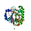
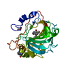
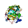
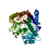
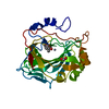
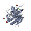
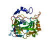
| Title | Exonuclease III homologue Mm3148 from Methanosarcina mazei | ||||||
 Components Components | Exodeoxyribonuclease III | ||||||
 Keywords Keywords | HYDROLASE / exonuclease III homologue / endonuclease / magnesium binding | ||||||
| Function / homology |  Function and homology information Function and homology information: / exodeoxyribonuclease III / endonuclease activity / DNA repair / DNA binding / metal ion binding Similarity search - Function | ||||||
| Biological species |  Methanosarcina mazei (archaea) Methanosarcina mazei (archaea) | ||||||
| Method |  X-RAY DIFFRACTION / SYNCHROTRON / MOLECULAR REPLACEMENT / molecular replacement / Resolution: 1.44 Å X-RAY DIFFRACTION / SYNCHROTRON / MOLECULAR REPLACEMENT / molecular replacement / Resolution: 1.44 Å | ||||||
 Authors Authors | Lakomek, K. / Dickmanns, A. / Ficner, R. | ||||||
 Citation Citation | Journal: To Be Published Title: Structure of the archael ExoIII homologue Mm3148 at 1.4 Angstrom Authors: Lakomek, K. / Dickmanns, A. / Ber, S. / Fritz, H.-J. / Ficner, R. | ||||||
| History |
|
- Structure visualization
| Structure viewer | Molecule: MolmilJmol/JSmol |
|---|
- Downloads & links
-Download
| PDBx/mmCIF format | 5j8n.cif.gz | 135.8 KB | Display | PDBx/mmCIF format |
|---|---|---|---|---|
| PDB format | pdb5j8n.ent.gz | 104.2 KB | Display | PDB format |
| PDBx/mmJSON format | 5j8n.json.gz | Tree view | PDBx/mmJSON format | |
| Others |  Other downloads Other downloads |
-Validation report
| Summary document | 5j8n_validation.pdf.gz | 425.2 KB | Display | wwPDB validaton report |
|---|---|---|---|---|
| Full document | 5j8n_full_validation.pdf.gz | 426.1 KB | Display | |
| Data in XML | 5j8n_validation.xml.gz | 15.1 KB | Display | |
| Data in CIF | 5j8n_validation.cif.gz | 22.8 KB | Display | |
| Arichive directory | https://data.pdbj.org/pub/pdb/validation_reports/j8/5j8nftp://data.pdbj.org/pub/pdb/validation_reports/j8/5j8n | HTTPS FTP |
-Related structure data
| Related structure data |  3fziS S: Starting model for refinement |
|---|---|
| Similar structure data |


-Links
 PDBj
PDBj

- Assembly
| Deposited unit | 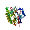
| ||||||||
|---|---|---|---|---|---|---|---|---|---|
| 1 |
| ||||||||
| Unit cell |
|
-Components
| #1: Protein | Mass: 29817.801 Da / Num. of mol.: 1 / Mutation: P2A, A118M, M209Y Source method: isolated from a genetically manipulated source Source: (gene. exp.) Methanosarcina mazei (archaea)Strain: ATCC BAA-159 / DSM 3647 / Goe1 / Go1 / JCM 11833 / OCM 88 Gene: MM_3148 / Plasmid: pET21-d derivative / Production host:  |
|---|---|
| #2: Chemical | ChemComp-MG /   Mass: 24.305 Da / Num. of mol.: 1 / Source method: obtained synthetically / Formula: Mg Mass: 24.305 Da / Num. of mol.: 1 / Source method: obtained synthetically / Formula: Mg |
| #3: Chemical | ChemComp-PG4 /   Mass: 194.226 Da / Num. of mol.: 1 / Source method: obtained synthetically / Formula: C8H18O5 / Comment: precipitant*YM Mass: 194.226 Da / Num. of mol.: 1 / Source method: obtained synthetically / Formula: C8H18O5 / Comment: precipitant*YM |
| #4: Water | ChemComp-HOH /  Mass: 18.015 Da / Num. of mol.: 317 / Source method: isolated from a natural source / Formula: H2O Mass: 18.015 Da / Num. of mol.: 317 / Source method: isolated from a natural source / Formula: H2O |
-Experimental details
-Experiment
| Experiment | Method: X-RAY DIFFRACTION / Number of used crystals: 1 |
|---|
- Sample preparation
| Crystal | Density Matthews: 2.15 Å3/Da / Density % sol: 42.72 % / Description: plate-like shape |
|---|---|
| Crystal grow | Temperature: 293 K / Method: vapor diffusion, sitting drop / pH: 8.5 Details: 20 % (w/v) PEG 8000, 20 % (w/v) PEG 400, 100 mM Tris/HCl pH 8.5, 100 mM MgCl2 mixed 1:1 with 10 mg/ml protein dissolved in 300 mM NaCl, 10 mM KHEPES pH 7.6, 2 mM DTT and addition of a seed ...Details: 20 % (w/v) PEG 8000, 20 % (w/v) PEG 400, 100 mM Tris/HCl pH 8.5, 100 mM MgCl2 mixed 1:1 with 10 mg/ml protein dissolved in 300 mM NaCl, 10 mM KHEPES pH 7.6, 2 mM DTT and addition of a seed stock solution (1/10 volume) |
-Data collection
| Diffraction | Mean temperature: 100 K | |||||||||||||||||||||||||||||||||||||||||||||||||||||||
|---|---|---|---|---|---|---|---|---|---|---|---|---|---|---|---|---|---|---|---|---|---|---|---|---|---|---|---|---|---|---|---|---|---|---|---|---|---|---|---|---|---|---|---|---|---|---|---|---|---|---|---|---|---|---|---|---|
| Diffraction source | Source: SYNCHROTRON / Site: EMBL/DESY, HAMBURG  / Beamline: X11 / Wavelength: 0.815 Å / Beamline: X11 / Wavelength: 0.815 Å | |||||||||||||||||||||||||||||||||||||||||||||||||||||||
| Detector | Type: MAR CCD 165 mm / Detector: CCD / Date: May 16, 2007 | |||||||||||||||||||||||||||||||||||||||||||||||||||||||
| Radiation | Monochromator: 0.99 / Protocol: SINGLE WAVELENGTH / Monochromatic (M) / Laue (L): M / Scattering type: x-ray | |||||||||||||||||||||||||||||||||||||||||||||||||||||||
| Radiation wavelength | Wavelength: 0.815 Å / Relative weight: 1 | |||||||||||||||||||||||||||||||||||||||||||||||||||||||
| Reflection | Resolution: 1.44→15 Å / Num. obs: 47242 / % possible obs: 99.9 % / Redundancy: 6.3 % / Biso Wilson estimate: 14.63 Å2 / Rmerge(I) obs: 0.052 / Net I/av σ(I): 32.105 / Net I/σ(I): 13.3 | |||||||||||||||||||||||||||||||||||||||||||||||||||||||
| Reflection shell |
|
-Phasing
| Phasing | Method: molecular replacement |
|---|
- Processing
| Software |
| ||||||||||||||||||||||||||||||||||||||||
|---|---|---|---|---|---|---|---|---|---|---|---|---|---|---|---|---|---|---|---|---|---|---|---|---|---|---|---|---|---|---|---|---|---|---|---|---|---|---|---|---|---|
| Refinement | Method to determine structure: MOLECULAR REPLACEMENT Starting model: 3FZI Resolution: 1.44→14.831 Å / SU ML: 0.09 / Cross valid method: FREE R-VALUE / σ(F): 1.34 / Phase error: 15.62 / Stereochemistry target values: ML
| ||||||||||||||||||||||||||||||||||||||||
| Solvent computation | Shrinkage radii: 0.9 Å / VDW probe radii: 1.11 Å / Solvent model: FLAT BULK SOLVENT MODEL | ||||||||||||||||||||||||||||||||||||||||
| Displacement parameters | Biso max: 76.67 Å2 / Biso mean: 21.2763 Å2 / Biso min: 9.48 Å2 | ||||||||||||||||||||||||||||||||||||||||
| Refinement step | Cycle: final / Resolution: 1.44→14.831 Å
| ||||||||||||||||||||||||||||||||||||||||
| Refinement TLS params. | Method: refined / Origin x: -17.8847 Å / Origin y: -2.734 Å / Origin z: 3.3491 Å
| ||||||||||||||||||||||||||||||||||||||||
| Refinement TLS group |
|
