 Movie
Movie Controller


+ Open data

- Basic information
| Entry | Database: PDB / ID: 5j6q | |||||||||
|---|---|---|---|---|---|---|---|---|---|---|
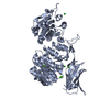
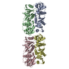
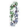
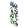
| Title | Cwp8 from Clostridium difficile | |||||||||
 Components Components | Cell wall binding protein cwp8 | |||||||||
 Keywords Keywords | CELL ADHESION / cell wall protein / S-layer / CWB2 domain / toprim fold | |||||||||
| Function / homology | : / : / Cell wall binding protein Cwp8 domain 2 / Cell wall binding protein Cwp8 domain 3 / : / Putative cell wall binding repeat 2 / Cell wall binding domain 2 (CWB2) / Cell wall binding protein cwp8 Function and homology information Function and homology information | |||||||||
| Biological species |  Peptoclostridium difficile (bacteria) Peptoclostridium difficile (bacteria) | |||||||||
| Method |  X-RAY DIFFRACTION / SYNCHROTRON / SAD / Resolution: 2.1 Å X-RAY DIFFRACTION / SYNCHROTRON / SAD / Resolution: 2.1 Å | |||||||||
 Authors Authors | Renko, M. / Usenik, A. / Turk, D. | |||||||||
| Funding support |  Slovenia, 2items Slovenia, 2items
| |||||||||
 Citation Citation | Journal: Structure / Year: 2017 Title: The CWB2 Cell Wall-Anchoring Module Is Revealed by the Crystal Structures of the Clostridium difficile Cell Wall Proteins Cwp8 and Cwp6. Authors: Usenik, A. / Renko, M. / Mihelic, M. / Lindic, N. / Borisek, J. / Perdih, A. / Pretnar, G. / Muller, U. / Turk, D. | |||||||||
| History |
|
- Structure visualization
| Structure viewer | Molecule: MolmilJmol/JSmol |
|---|
- Downloads & links
-Download
| PDBx/mmCIF format | 5j6q.cif.gz | 246.8 KB | Display | PDBx/mmCIF format |
|---|---|---|---|---|
| PDB format | pdb5j6q.ent.gz | 198.5 KB | Display | PDB format |
| PDBx/mmJSON format | 5j6q.json.gz | Tree view | PDBx/mmJSON format | |
| Others |  Other downloads Other downloads |
-Validation report
| Arichive directory | https://data.pdbj.org/pub/pdb/validation_reports/j6/5j6qftp://data.pdbj.org/pub/pdb/validation_reports/j6/5j6q | HTTPS FTP |
|---|
-Related structure data


-Links
 PDBj
PDBj- Assembly
| Deposited unit | 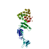
| ||||||||
|---|---|---|---|---|---|---|---|---|---|
| 1 |
| ||||||||
| Unit cell |
|
-Components
| #1: Protein | Mass: 64816.691 Da / Num. of mol.: 1 Source method: isolated from a genetically manipulated source Details: genomic DNA from C. difficile 630 Source: (gene. exp.) Peptoclostridium difficile (strain 630) (bacteria)Gene: cwp8, CD630_27990 / Plasmid: pMCSG7 / Production host: | ||||
|---|---|---|---|---|---|
| #2: Chemical | ChemComp-SO4 /   Mass: 96.063 Da / Num. of mol.: 8 / Source method: obtained synthetically / Formula: SO4 Mass: 96.063 Da / Num. of mol.: 8 / Source method: obtained synthetically / Formula: SO4#3: Chemical | ChemComp-CL / |   Mass: 35.453 Da / Num. of mol.: 1 / Source method: obtained synthetically / Formula: Cl Mass: 35.453 Da / Num. of mol.: 1 / Source method: obtained synthetically / Formula: Cl#4: Water | ChemComp-HOH / |  Mass: 18.015 Da / Num. of mol.: 412 / Source method: isolated from a natural source / Formula: H2O Mass: 18.015 Da / Num. of mol.: 412 / Source method: isolated from a natural source / Formula: H2O |
-Experimental details
-Experiment
| Experiment | Method: X-RAY DIFFRACTION / Number of used crystals: 2 |
|---|
- Sample preparation
| Crystal |
| ||||||||||||
|---|---|---|---|---|---|---|---|---|---|---|---|---|---|
| Crystal grow |
|
-Data collection
| Diffraction |
| ||||||||||||||||||||||||
|---|---|---|---|---|---|---|---|---|---|---|---|---|---|---|---|---|---|---|---|---|---|---|---|---|---|
| Diffraction source |
| ||||||||||||||||||||||||
| Detector |
| ||||||||||||||||||||||||
| Radiation |
| ||||||||||||||||||||||||
| Radiation wavelength |
| ||||||||||||||||||||||||
| Reflection | Entry-ID: 5J6Q / % possible obs: 95 %
| ||||||||||||||||||||||||
| Reflection shell |
|


- Processing
| Software |
| |||||||||||||||||||||||||
|---|---|---|---|---|---|---|---|---|---|---|---|---|---|---|---|---|---|---|---|---|---|---|---|---|---|---|
| Refinement | Method to determine structure: SAD / Resolution: 2.1→42.09 Å / Cor.coef. Fo:Fc: 0.9451 / Cor.coef. Fo:Fc free: 0.9351 / Cross valid method: NONE / σ(F): 0 / Phase error: 27.2 Details: Free Kick ML target function uses all data for calculation of phase error estimates from randomly displaced atoms. Praznikar, J. & Turk, D. (2014) Free kick instead of cross-validation in ...Details: Free Kick ML target function uses all data for calculation of phase error estimates from randomly displaced atoms. Praznikar, J. & Turk, D. (2014) Free kick instead of cross-validation in maximum-likelihood refinement of macromolecular crystal structures. Acta Cryst. D70, 3124-3134.
| |||||||||||||||||||||||||
| Solvent computation | VDW probe radii: 1.6 Å / Bsol: 28.69 Å2 / ksol: 0.35 e/Å3 | |||||||||||||||||||||||||
| Displacement parameters | Biso max: 199.2 Å2 / Biso mean: 74.43 Å2 / Biso min: 20.06 Å2
| |||||||||||||||||||||||||
| Refinement step | Cycle: LAST / Resolution: 2.1→42.09 Å
| |||||||||||||||||||||||||
| LS refinement shell | Resolution: 2.1→2.135 Å
|
