 Movie
Movie Controller

[English] 日本語
 Yorodumi
- PDB-5j4n: Crystal structure of the L-arginine/agmatine antiporter AdiC in c... -
+ Open data

- Basic information
| Entry | Database: PDB / ID: 5j4n | ||||||
|---|---|---|---|---|---|---|---|
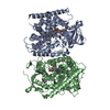
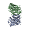
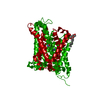
| Title | Crystal structure of the L-arginine/agmatine antiporter AdiC in complex with agmatine at 2.6 Angstroem resolution | ||||||
 Components Components | Arginine/agmatine antiporter | ||||||
 Keywords Keywords | TRANSPORT PROTEIN / Membrane Protein / Exchanger / Transporter / AdiC-agmatine complex | ||||||
| Function / homology |  Function and homology information Function and homology informationarginine:agmatine antiporter activity / cellular stress response to acidic pH / antiporter activity / amino acid transport / identical protein binding / plasma membrane Similarity search - Function | ||||||
| Biological species |  | ||||||
| Method |  X-RAY DIFFRACTION / SYNCHROTRON / MOLECULAR REPLACEMENT / Resolution: 2.594 Å X-RAY DIFFRACTION / SYNCHROTRON / MOLECULAR REPLACEMENT / Resolution: 2.594 Å | ||||||
 Authors Authors | Jeckelmann, J.M. / Ilgue, H. / Fotiadis, D. | ||||||
| Funding support |  Switzerland, 1items Switzerland, 1items
| ||||||
 Citation Citation | Journal: Proc.Natl.Acad.Sci.USA / Year: 2016 Title: Insights into the molecular basis for substrate binding and specificity of the wild-type L-arginine/agmatine antiporter AdiC. Authors: Ilgu, H. / Jeckelmann, J.M. / Gapsys, V. / Ucurum, Z. / de Groot, B.L. / Fotiadis, D. | ||||||
| History |
|
- Structure visualization
| Structure viewer | Molecule: MolmilJmol/JSmol |
|---|
- Downloads & links
-Download
| PDBx/mmCIF format | 5j4n.cif.gz | 170.9 KB | Display | PDBx/mmCIF format |
|---|---|---|---|---|
| PDB format | pdb5j4n.ent.gz | 135.8 KB | Display | PDB format |
| PDBx/mmJSON format | 5j4n.json.gz | Tree view | PDBx/mmJSON format | |
| Others |  Other downloads Other downloads |
-Validation report
| Arichive directory | https://data.pdbj.org/pub/pdb/validation_reports/j4/5j4nftp://data.pdbj.org/pub/pdb/validation_reports/j4/5j4n | HTTPS FTP |
|---|
-Related structure data
| Related structure data |  5j4iSC S: Starting model for refinement C: citing same article ( |
|---|---|
| Similar structure data |


-Links
 PDBj
PDBj- Assembly
| Deposited unit | 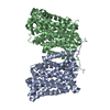
| ||||||||
|---|---|---|---|---|---|---|---|---|---|
| 1 |
| ||||||||
| Unit cell |
|
-Components
| #1: Protein | Mass: 47841.387 Da / Num. of mol.: 2 Source method: isolated from a genetically manipulated source Details: in complex with agmatine / Source: (gene. exp.) #2: Chemical |   Mass: 130.191 Da / Num. of mol.: 2 / Source method: obtained synthetically / Formula: C5H14N4 Mass: 130.191 Da / Num. of mol.: 2 / Source method: obtained synthetically / Formula: C5H14N4#3: Water | ChemComp-HOH / |  Mass: 18.015 Da / Num. of mol.: 12 / Source method: isolated from a natural source / Formula: H2O Mass: 18.015 Da / Num. of mol.: 12 / Source method: isolated from a natural source / Formula: H2O |
|---|
-Experimental details
-Experiment
| Experiment | Method: X-RAY DIFFRACTION / Number of used crystals: 1 |
|---|
- Sample preparation
| Crystal | Density Matthews: 3.35 Å3/Da / Density % sol: 63.3 % |
|---|---|
| Crystal grow | Temperature: 298 K / Method: vapor diffusion, sitting drop / pH: 7 / Details: PEG 400; NaCl |
-Data collection
| Diffraction | Mean temperature: 100 K |
|---|---|
| Diffraction source | Source: SYNCHROTRON / Site: SLS / Beamline: X06SA / Wavelength: 1 Å |
| Detector | Type: DECTRIS PILATUS3 6M / Detector: PIXEL / Date: Aug 23, 2015 |
| Radiation | Protocol: SINGLE WAVELENGTH / Monochromatic (M) / Laue (L): M / Scattering type: x-ray |
| Radiation wavelength | Wavelength: 1 Å / Relative weight: 1 |
| Reflection | Resolution: 2.594→49.44 Å / Num. obs: 41837 / % possible obs: 98.7 % / Redundancy: 6.7 % / CC1/2: 0.999 / Rmerge(I) obs: 0.0109 / Net I/σ(I): 12 |
| Reflection shell | Resolution: 2.594→2.73 Å / Redundancy: 6.7 % / Rmerge(I) obs: 1.17 / Mean I/σ(I) obs: 1.6 / % possible all: 94.9 |
- Processing
| Software |
| |||||||||||||||||||||||||||||||||||||||||||||||||||||||||||||||||||||||||||||||||||||||||||||||||||||||||||||||||||||||||||||||||||||||||||||||||||||||||||||||||||||||||||||||||||||||||||||||||||||||||||
|---|---|---|---|---|---|---|---|---|---|---|---|---|---|---|---|---|---|---|---|---|---|---|---|---|---|---|---|---|---|---|---|---|---|---|---|---|---|---|---|---|---|---|---|---|---|---|---|---|---|---|---|---|---|---|---|---|---|---|---|---|---|---|---|---|---|---|---|---|---|---|---|---|---|---|---|---|---|---|---|---|---|---|---|---|---|---|---|---|---|---|---|---|---|---|---|---|---|---|---|---|---|---|---|---|---|---|---|---|---|---|---|---|---|---|---|---|---|---|---|---|---|---|---|---|---|---|---|---|---|---|---|---|---|---|---|---|---|---|---|---|---|---|---|---|---|---|---|---|---|---|---|---|---|---|---|---|---|---|---|---|---|---|---|---|---|---|---|---|---|---|---|---|---|---|---|---|---|---|---|---|---|---|---|---|---|---|---|---|---|---|---|---|---|---|---|---|---|---|---|---|---|---|---|---|
| Refinement | Method to determine structure: MOLECULAR REPLACEMENT Starting model: 5J4I Resolution: 2.594→49.44 Å / SU ML: 0.33 / Cross valid method: FREE R-VALUE / σ(F): 1.31 / Phase error: 26.51
| |||||||||||||||||||||||||||||||||||||||||||||||||||||||||||||||||||||||||||||||||||||||||||||||||||||||||||||||||||||||||||||||||||||||||||||||||||||||||||||||||||||||||||||||||||||||||||||||||||||||||||
| Solvent computation | Shrinkage radii: 0.9 Å / VDW probe radii: 1.11 Å | |||||||||||||||||||||||||||||||||||||||||||||||||||||||||||||||||||||||||||||||||||||||||||||||||||||||||||||||||||||||||||||||||||||||||||||||||||||||||||||||||||||||||||||||||||||||||||||||||||||||||||
| Displacement parameters | Biso mean: 68.8 Å2 | |||||||||||||||||||||||||||||||||||||||||||||||||||||||||||||||||||||||||||||||||||||||||||||||||||||||||||||||||||||||||||||||||||||||||||||||||||||||||||||||||||||||||||||||||||||||||||||||||||||||||||
| Refinement step | Cycle: LAST / Resolution: 2.594→49.44 Å
| |||||||||||||||||||||||||||||||||||||||||||||||||||||||||||||||||||||||||||||||||||||||||||||||||||||||||||||||||||||||||||||||||||||||||||||||||||||||||||||||||||||||||||||||||||||||||||||||||||||||||||
| Refine LS restraints |
| |||||||||||||||||||||||||||||||||||||||||||||||||||||||||||||||||||||||||||||||||||||||||||||||||||||||||||||||||||||||||||||||||||||||||||||||||||||||||||||||||||||||||||||||||||||||||||||||||||||||||||
| LS refinement shell |
|
