 Movie
Movie Controller

[English] 日本語
 Yorodumi
- PDB-5j1b: structure of the core domaine of Knr4, an intrinsically disordere... -
+ Open data

- Basic information
| Entry | Database: PDB / ID: 5j1b | ||||||
|---|---|---|---|---|---|---|---|
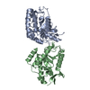
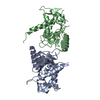
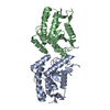
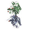
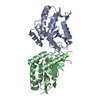
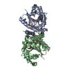
| Title | structure of the core domaine of Knr4, an intrinsically disordered protein from Saccharomyces cerevisiae - WT. | ||||||
 Components Components | Cell wall assembly regulator SMI1 | ||||||
 Keywords Keywords | SIGNALING PROTEIN / cell-wall integrity / stress response / beta-glucan synthesis | ||||||
| Function / homology |  Function and homology information Function and homology informationregulation of fungal-type cell wall biogenesis / fungal-type cell wall beta-glucan biosynthetic process / incipient cellular bud site / cellular bud neck / mating projection tip / regulation of mitotic cell cycle / cell wall organization / DNA binding / nucleus Similarity search - Function | ||||||
| Biological species |  | ||||||
| Method |  X-RAY DIFFRACTION / SYNCHROTRON / SAD / Resolution: 2.5 Å X-RAY DIFFRACTION / SYNCHROTRON / SAD / Resolution: 2.5 Å | ||||||
 Authors Authors | Maveyraud, L. / Batista, M. / Martin-yken, H. / Francois, J.M. / Zerbib, D. / Mourey, L. | ||||||
 Citation Citation | Journal: To Be Published Title: Structural and biophysical characterization of Knr4, an intrinsically disordered hub protein form Saccharomyces cerevisiae Authors: Batista, M. / Maveyraud, L. / Zerbib, D. | ||||||
| History |
|
- Structure visualization
| Structure viewer | Molecule: MolmilJmol/JSmol |
|---|
- Downloads & links
-Download
| PDBx/mmCIF format | 5j1b.cif.gz | 184.7 KB | Display | PDBx/mmCIF format |
|---|---|---|---|---|
| PDB format | pdb5j1b.ent.gz | 148.1 KB | Display | PDB format |
| PDBx/mmJSON format | 5j1b.json.gz | Tree view | PDBx/mmJSON format | |
| Others |  Other downloads Other downloads |
-Validation report
| Summary document | 5j1b_validation.pdf.gz | 436.9 KB | Display | wwPDB validaton report |
|---|---|---|---|---|
| Full document | 5j1b_full_validation.pdf.gz | 438 KB | Display | |
| Data in XML | 5j1b_validation.xml.gz | 15.9 KB | Display | |
| Data in CIF | 5j1b_validation.cif.gz | 21 KB | Display | |
| Arichive directory | https://data.pdbj.org/pub/pdb/validation_reports/j1/5j1bftp://data.pdbj.org/pub/pdb/validation_reports/j1/5j1b | HTTPS FTP |
-Related structure data
| Similar structure data |
|---|


-Links
 PDBj
PDBj- Assembly
| Deposited unit | 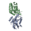
| ||||||||
|---|---|---|---|---|---|---|---|---|---|
| 1 |
| ||||||||
| Unit cell |
|
-Components
| #1: Protein | Mass: 30921.141 Da / Num. of mol.: 2 / Fragment: UNP residues 80-340 Source method: isolated from a genetically manipulated source Source: (gene. exp.) Gene: SMI1, KNR4, KTR4, YGR229C, G8553 / Plasmid: pGEX-6P3 / Production host:  |
|---|
-Experimental details
-Experiment
| Experiment | Method: X-RAY DIFFRACTION / Number of used crystals: 1 |
|---|
- Sample preparation
| Crystal | Density Matthews: 2.85 Å3/Da / Density % sol: 56.9 % |
|---|---|
| Crystal grow | Temperature: 285 K / Method: vapor diffusion, hanging drop / Details: PEG (3000-6000) 15 - 24 % (w/v) pH 8.0 - 9.0 / PH range: 8.0-9.0 |
-Data collection
| Diffraction | Mean temperature: 100 K |
|---|---|
| Diffraction source | Source: SYNCHROTRON / Site: ESRF  / Beamline: ID23-1 / Wavelength: 0.97942 Å / Beamline: ID23-1 / Wavelength: 0.97942 Å |
| Detector | Type: DECTRIS PILATUS 6M / Detector: PIXEL / Date: Jan 15, 2015 |
| Radiation | Protocol: SINGLE WAVELENGTH / Monochromatic (M) / Laue (L): M / Scattering type: x-ray |
| Radiation wavelength | Wavelength: 0.97942 Å / Relative weight: 1 |
| Reflection | Resolution: 2.5→40.25 Å / Num. obs: 19048 / % possible obs: 99.7 % / Redundancy: 2.6 % / Biso Wilson estimate: 82 Å2 / CC1/2: 0.999 / Rsym value: 0.04 / Net I/σ(I): 12.7 |
| Reflection shell | Resolution: 2.5→2.65 Å / Redundancy: 2.6 % / Rmerge(I) obs: 0.908 / Mean I/σ(I) obs: 1 / % possible all: 99.7 |
- Processing
| Software |
| ||||||||||||||||||||||||||||||||||||||||||||||||||||||||||||||||||||||||||||||||||||||||||||||||||||||||||||||||||||||||||||||||||||||||||||||||||||||||||||||||||||||||||||||||||||||
|---|---|---|---|---|---|---|---|---|---|---|---|---|---|---|---|---|---|---|---|---|---|---|---|---|---|---|---|---|---|---|---|---|---|---|---|---|---|---|---|---|---|---|---|---|---|---|---|---|---|---|---|---|---|---|---|---|---|---|---|---|---|---|---|---|---|---|---|---|---|---|---|---|---|---|---|---|---|---|---|---|---|---|---|---|---|---|---|---|---|---|---|---|---|---|---|---|---|---|---|---|---|---|---|---|---|---|---|---|---|---|---|---|---|---|---|---|---|---|---|---|---|---|---|---|---|---|---|---|---|---|---|---|---|---|---|---|---|---|---|---|---|---|---|---|---|---|---|---|---|---|---|---|---|---|---|---|---|---|---|---|---|---|---|---|---|---|---|---|---|---|---|---|---|---|---|---|---|---|---|---|---|---|---|
| Refinement | Method to determine structure: SAD / Resolution: 2.5→35 Å / Cor.coef. Fo:Fc: 0.968 / Cor.coef. Fo:Fc free: 0.956 / SU B: 29.377 / SU ML: 0.259 / Cross valid method: THROUGHOUT / ESU R: 0.418 / ESU R Free: 0.253 / Stereochemistry target values: MAXIMUM LIKELIHOOD / Details: HYDROGENS HAVE BEEN ADDED IN THE RIDING POSITIONS
| ||||||||||||||||||||||||||||||||||||||||||||||||||||||||||||||||||||||||||||||||||||||||||||||||||||||||||||||||||||||||||||||||||||||||||||||||||||||||||||||||||||||||||||||||||||||
| Solvent computation | Ion probe radii: 0.8 Å / Shrinkage radii: 0.8 Å / VDW probe radii: 1.1 Å / Solvent model: MASK | ||||||||||||||||||||||||||||||||||||||||||||||||||||||||||||||||||||||||||||||||||||||||||||||||||||||||||||||||||||||||||||||||||||||||||||||||||||||||||||||||||||||||||||||||||||||
| Displacement parameters | Biso mean: 85.129 Å2
| ||||||||||||||||||||||||||||||||||||||||||||||||||||||||||||||||||||||||||||||||||||||||||||||||||||||||||||||||||||||||||||||||||||||||||||||||||||||||||||||||||||||||||||||||||||||
| Refinement step | Cycle: LAST / Resolution: 2.5→35 Å
| ||||||||||||||||||||||||||||||||||||||||||||||||||||||||||||||||||||||||||||||||||||||||||||||||||||||||||||||||||||||||||||||||||||||||||||||||||||||||||||||||||||||||||||||||||||||
| Refine LS restraints |
|