Redundancy: 7.1 % / Av σ(I) over netI: 27.44 / Number: 224942 / Rmerge(I) obs: 0.062 / Χ2: 0.89 / D res high: 2.1 Å / D res low: 50 Å / Num. obs: 31606 / % possible obs: 99.8
D res high: 2.1 Å / D res low: 50 Å / FOM : 0.347 / FOM acentric: 0.371 / FOM centric: 0.164 / Reflection: 30355 / Reflection acentric: 26744 / Reflection centric: 3611
Phasing MAD set
Highest resolution: 2.1 Å / Lowest resolution: 50 Å
ID
R cullis acentric
R cullis centric
Loc acentric
Loc centric
Power acentric
Power centric
Reflection acentric
Reflection centric
1
1.49
1
0.3
0.3
0
0
26744
3611
2
0.97
0.94
37.7
53.2
0.38
0.31
17064
2636
Phasing MAD set shell
ID
Resolution (Å)
R cullis acentric
R cullis centric
Loc acentric
Loc centric
Power acentric
Power centric
Reflection acentric
Reflection centric
1
12.98-50
1.05
1
0.7
0.6
0
0
72
73
1
7.46-12.98
1.14
1
0.9
0.7
0
0
419
182
1
5.23-7.46
1.57
1
0.7
0.4
0
0
1085
304
1
4.03-5.23
1.07
1
0.6
0.5
0
0
2045
419
1
3.28-4.03
1.2
1
0.4
0.3
0
0
3289
524
1
2.76-3.28
1.89
1
0.4
0.2
0
0
4866
643
1
2.39-2.76
2.25
1
0.2
0.1
0
0
6726
746
1
2.1-2.39
1.62
1
0.1
0
0
0
8242
720
2
12.98-50
0.92
0.9
55.6
60.8
0.71
0.63
68
68
2
7.46-12.98
0.91
0.89
54.9
57.3
0.61
0.55
417
178
2
5.23-7.46
0.93
0.87
38
47.6
0.74
0.55
1085
304
2
4.03-5.23
0.96
0.94
50.1
66.2
0.45
0.32
2045
419
2
3.28-4.03
0.98
0.95
44.6
62.5
0.36
0.24
3289
524
2
2.76-3.28
0.98
0.96
33.5
45.7
0.35
0.22
4866
643
2
2.39-2.76
0.99
0.98
30.8
42.9
0.26
0.15
5294
500
2
2.1-2.39
0
0
0
0
0
0
0
0
Phasing MAD set site
Atom type symbol: Se
ID
B iso
Fract x
Fract y
Fract z
Occupancy
Occupancy iso
1
21.7385
0.207
0.4
0.142
7.337
0
2
27.1598
0.037
0.369
0.187
8.417
0
3
19.1715
-0.322
0.409
0.172
6.156
0
4
22.9331
-0.151
0.447
0.131
6.679
0
5
29.882
0.257
0.4
0.017
5.067
0
6
21.0161
-0.438
0.261
0.183
4.799
0
7
34.8677
-0.312
0.247
0.161
4.382
0
8
23.8419
-0.214
0.283
0.242
4.498
0
9
33.9147
0.084
0.492
0.046
4.093
0
10
41.0733
0.122
0.384
0.015
3.72
0
11
39.1673
-0.367
0.156
0.38
4.386
0
12
66.7456
0.272
0.393
-0.095
2.032
0
13
15.7467
0.206
0.399
0.141
4.358
-0.192
14
23.0023
0.037
0.368
0.188
4.795
-0.243
15
16.7049
-0.324
0.407
0.172
3.432
-0.17
16
23.6521
-0.152
0.447
0.132
4.206
-0.226
17
41.5724
0.255
0.398
0.017
2.284
-0.276
18
22.3385
-0.437
0.261
0.18
2.438
-0.243
19
31.451
-0.313
0.247
0.161
2.52
-0.168
20
15.7014
-0.212
0.282
0.241
2.916
-0.114
21
43.6228
0.081
0.492
0.046
3.041
-0.231
22
101.4886
0.124
0.384
0.016
3.095
-0.206
23
39.662
-0.362
0.153
0.382
2.698
-0.193
24
51.364
0.266
0.392
-0.096
1.045
-0.116
Phasing MAD shell
Resolution (Å)
FOM
FOM acentric
FOM centric
Reflection
Reflection acentric
Reflection centric
12.98-50
0.443
0.574
0.314
145
72
73
7.46-12.98
0.539
0.642
0.303
601
419
182
5.23-7.46
0.623
0.696
0.364
1389
1085
304
4.03-5.23
0.548
0.609
0.249
2464
2045
419
3.28-4.03
0.535
0.585
0.221
3813
3289
524
2.76-3.28
0.484
0.523
0.193
5509
4866
643
2.39-2.76
0.305
0.33
0.08
7472
6726
746
2.1-2.39
0.105
0.114
0
8962
8242
720
Phasing dm
Method: Solvent flattening and Histogram matching / Reflection: 30355
Phasing dm shell
Resolution (Å)
Delta phi final
FOM
Reflection
8.43-100
64.6
0.52
508
6.74-8.43
51.8
0.866
506
5.8-6.74
47.7
0.893
566
5.17-5.8
43.8
0.916
639
4.71-5.17
49.4
0.917
694
4.35-4.71
48.6
0.936
766
4.06-4.35
55
0.922
816
3.83-4.06
52.9
0.923
848
3.63-3.83
50.7
0.919
914
3.46-3.63
52
0.908
964
3.31-3.46
57.1
0.9
995
3.18-3.31
54.2
0.899
1031
3.06-3.18
56.5
0.884
1077
2.95-3.06
53.9
0.885
1109
2.86-2.95
56.4
0.889
1135
2.77-2.86
59.3
0.867
1178
2.69-2.77
61
0.854
1230
2.62-2.69
62.5
0.843
1240
2.55-2.62
64.9
0.808
1279
2.49-2.55
64.6
0.823
1300
2.44-2.49
70.1
0.83
1378
2.38-2.44
72.6
0.832
1351
2.33-2.38
75.1
0.811
1387
2.28-2.33
81
0.818
1428
2.24-2.28
78.5
0.813
1375
2.2-2.24
82.4
0.813
1414
2.16-2.2
83
0.774
1324
2.1-2.16
86
0.739
1903
-
Processing
Software
Name
Version
Classification
NB
DENZO
datareduction
SCALEPACK
datascaling
MLPHARE
phasing
DM
6.1
phasing
REFMAC
refinement
PDB_EXTRACT
3.1
dataextraction
SBC-Collect
datacollection
HKL-3000
datareduction
HKL-3000
datascaling
HKL-3000
phasing
SHELXD
phasing
SHELXE
modelbuilding
SOLVE
phasing
RESOLVE
phasing
ARP/wARP
modelbuilding
CCP4
phasing
O
modelbuilding
COO
phasing
Refinement
Method to determine structure: MAD / Resolution: 2.1→37.84 Å / Cor.coef. Fo:Fc: 0.958 / Cor.coef. Fo:Fc free: 0.935 / WRfactor Rfree: 0.2122 / WRfactor Rwork: 0.1592 / Occupancy max: 1 / Occupancy min: 0.4 / FOM work R set: 0.9134 / SU B: 7.167 / SU ML: 0.096 / SU R Cruickshank DPI: 0.2045 / SU Rfree: 0.17 / Cross valid method: THROUGHOUT / σ(F): 0 / ESU R Free: 0.17 Stereochemistry target values: MAXIMUM LIKELIHOOD WITH PHASES Details: HYDROGENS HAVE BEEN ADDED IN THE RIDING POSITIONS U VALUES : WITH TLS ADDED
Rfactor
Num. reflection
% reflection
Selection details
Rfree
0.2059
1518
5 %
RANDOM
Rwork
0.1594
-
-
-
all
0.1617
30241
-
-
obs
0.1617
30241
96.88 %
-
Solvent computation
Ion probe radii: 0.8 Å / Shrinkage radii: 0.8 Å / VDW probe radii: 1.2 Å / Solvent model: MASK
In the structure databanks used in Yorodumi, some data are registered as the other names, "COVID-19 virus" and "2019-nCoV". Here are the details of the virus and the list of structure data.
Jan 31, 2019. EMDB accession codes are about to change! (news from PDBe EMDB page)
EMDB accession codes are about to change! (news from PDBe EMDB page)
The allocation of 4 digits for EMDB accession codes will soon come to an end. Whilst these codes will remain in use, new EMDB accession codes will include an additional digit and will expand incrementally as the available range of codes is exhausted. The current 4-digit format prefixed with “EMD-” (i.e. EMD-XXXX) will advance to a 5-digit format (i.e. EMD-XXXXX), and so on. It is currently estimated that the 4-digit codes will be depleted around Spring 2019, at which point the 5-digit format will come into force.
The EM Navigator/Yorodumi systems omit the EMD- prefix.
Related info.:Q: What is EMD? / ID/Accession-code notation in Yorodumi/EM Navigator
Yorodumi is a browser for structure data from EMDB, PDB, SASBDB, etc.
This page is also the successor to EM Navigator detail page, and also detail information page/front-end page for Omokage search.
The word "yorodu" (or yorozu) is an old Japanese word meaning "ten thousand". "mi" (miru) is to see.
Related info.:EMDB / PDB / SASBDB / Comparison of 3 databanks / Yorodumi Search / Aug 31, 2016. New EM Navigator & Yorodumi / Yorodumi Papers / Jmol/JSmol / Function and homology information / Changes in new EM Navigator and Yorodumi
 Movie
Movie Controller

 Yorodumi
 Open data
 Basic information
 Components
 Keywords
 Function and homology information
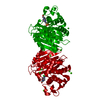
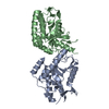
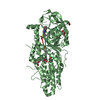
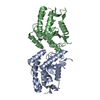
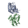
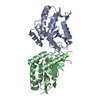
 Anaerococcus prevotii (bacteria)
 X-RAY DIFFRACTION /
  Authors
 Citation
 Structure visualization
 Downloads & links
 Other downloads


 PDBj
PDBj

 Assembly
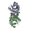

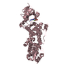
 Mass: 62.068 Da / Num. of mol.: 7 / Source method: obtained synthetically / Formula: C2H6O2
 Mass: 192.124 Da / Num. of mol.: 1 / Source method: obtained synthetically / Formula: C6H8O7
 Mass: 18.015 Da / Num. of mol.: 263 / Source method: isolated from a natural source / Formula: H2O
 Sample preparation
 / Beamline: 19-ID / Wavelength: 0.97918, 0.97929
 Processing