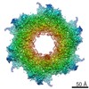
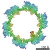
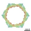
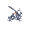
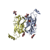
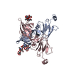
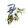
Journal: Nature / Year: 2016 Title: Structure of the T4 baseplate and its function in triggering sheath contraction. Authors: Nicholas M I Taylor / Nikolai S Prokhorov / Ricardo C Guerrero-Ferreira / Mikhail M Shneider / Christopher Browning / Kenneth N Goldie / Henning Stahlberg / Petr G Leiman / Abstract: Several systems, including contractile tail bacteriophages, the type VI secretion system and R-type pyocins, use a multiprotein tubular apparatus to attach to and penetrate host cell membranes. This ...Several systems, including contractile tail bacteriophages, the type VI secretion system and R-type pyocins, use a multiprotein tubular apparatus to attach to and penetrate host cell membranes. This macromolecular machine resembles a stretched, coiled spring (or sheath) wound around a rigid tube with a spike-shaped protein at its tip. A baseplate structure, which is arguably the most complex part of this assembly, relays the contraction signal to the sheath. Here we present the atomic structure of the approximately 6-megadalton bacteriophage T4 baseplate in its pre- and post-host attachment states and explain the events that lead to sheath contraction in atomic detail. We establish the identity and function of a minimal set of components that is conserved in all contractile injection systems and show that the triggering mechanism is universally conserved.
Monochromator: SI(111) MONOCHROMATOR / Protocol: SINGLE WAVELENGTH / Monochromatic (M) / Laue (L): M / Scattering type: x-ray
Radiation wavelength
Wavelength: 0.9793 Å / Relative weight: 1
Reflection
Resolution: 2.47→74.06 Å / Num. obs: 20716 / % possible obs: 100 % / Redundancy: 23.4 % / Biso Wilson estimate: 51.75 Å2 / CC1/2: 0.999 / Rmerge(I) obs: 0.085 / Net I/av σ(I): 30.8 / Net I/σ(I): 30.8
Reflection shell
Resolution (Å)
Redundancy (%)
Rmerge(I) obs
Mean I/σ(I) obs
Diffraction-ID
% possible all
2.47-2.57
24.8
0.843
5.2
1
100
8.91-74.06
19.1
0.043
1
99.5
-
Processing
Software
Name
Version
Classification
PHENIX
(dev_2328: ???)
refinement
SCALA
0.5.23
datascaling
PDB_EXTRACT
3.2
dataextraction
iMOSFLM
7.2.1
datareduction
SHELXCD
2006/3
phasing
Refinement
Method to determine structure: SAD / Resolution: 2.47→69.86 Å / SU ML: 0.3 / Cross valid method: THROUGHOUT / σ(F): 0.63 / Phase error: 20.17 / Stereochemistry target values: ML
Rfactor
Num. reflection
% reflection
Selection details
Rfree
0.2172
1774
4.71 %
random
Rwork
0.1898
-
-
-
obs
0.1911
20649
99.93 %
-
Solvent computation
Shrinkage radii: 0.9 Å / VDW probe radii: 1.11 Å / Solvent model: FLAT BULK SOLVENT MODEL
 Movie
Movie Controller

 Yorodumi
 Open data
 Basic information
 Components
 Keywords
 Function and homology information
 Enterobacteria phage T4 (virus)
 X-RAY DIFFRACTION /
  Authors
 Switzerland, 1items
  Citation
 Structure visualization
 Downloads & links
 Other downloads
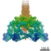


 PDBj
PDBj Assembly
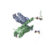

 Mass: 18.015 Da / Num. of mol.: 104 / Source method: isolated from a natural source / Formula: H2O
 Sample preparation
 Processing