 Movie
Movie Controller

[English] 日本語
 Yorodumi
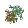
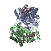
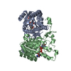
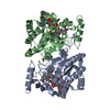
- PDB-5itv: Crystal structure of Bacillus subtilis BacC Dihydroanticapsin 7-d... -
+ Open data

- Basic information
| Entry | Database: PDB / ID: 5itv | ||||||
|---|---|---|---|---|---|---|---|
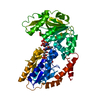
| Title | Crystal structure of Bacillus subtilis BacC Dihydroanticapsin 7-dehydrogenase in complex with NADH | ||||||
 Components Components | Dihydroanticapsin 7-dehydrogenase | ||||||
 Keywords Keywords | OXIDOREDUCTASE / short-chain dehydrogenases/reductases / Rossmann fold / NAD(P) binding domain | ||||||
| Function / homology |  Function and homology information Function and homology informationdihydroanticapsin dehydrogenase / bile acid metabolic process / antibiotic biosynthetic process / oxidoreductase activity Similarity search - Function | ||||||
| Biological species |  | ||||||
| Method |  X-RAY DIFFRACTION / MOLECULAR REPLACEMENT / Resolution: 2.26 Å X-RAY DIFFRACTION / MOLECULAR REPLACEMENT / Resolution: 2.26 Å | ||||||
 Authors Authors | Perinbam, K. / Balaram, H. / Row, T.N.G. / Gopal, B. | ||||||
 Citation Citation | Journal: Protein Eng. Des. Sel. / Year: 2017 Title: Probing the influence of non-covalent contact networks identified by charge density analysis on the oxidoreductase BacC. Authors: Perinbam, K. / Balaram, H. / Guru Row, T.N. / Gopal, B. | ||||||
| History |
|
- Structure visualization
| Structure viewer | Molecule: MolmilJmol/JSmol |
|---|
- Downloads & links
-Download
| PDBx/mmCIF format | 5itv.cif.gz | 197.1 KB | Display | PDBx/mmCIF format |
|---|---|---|---|---|
| PDB format | pdb5itv.ent.gz | 158.9 KB | Display | PDB format |
| PDBx/mmJSON format | 5itv.json.gz | Tree view | PDBx/mmJSON format | |
| Others |  Other downloads Other downloads |
-Validation report
| Arichive directory | https://data.pdbj.org/pub/pdb/validation_reports/it/5itvftp://data.pdbj.org/pub/pdb/validation_reports/it/5itv | HTTPS FTP |
|---|
-Related structure data


-Links
 PDBj
PDBj


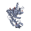
- Assembly
| Deposited unit | 
| ||||||||||||||||||||||||||||||||||||||||||||||||||||||||||||||||||||||||||||||||||||||||||||||||||
|---|---|---|---|---|---|---|---|---|---|---|---|---|---|---|---|---|---|---|---|---|---|---|---|---|---|---|---|---|---|---|---|---|---|---|---|---|---|---|---|---|---|---|---|---|---|---|---|---|---|---|---|---|---|---|---|---|---|---|---|---|---|---|---|---|---|---|---|---|---|---|---|---|---|---|---|---|---|---|---|---|---|---|---|---|---|---|---|---|---|---|---|---|---|---|---|---|---|---|---|
| 1 | 
| ||||||||||||||||||||||||||||||||||||||||||||||||||||||||||||||||||||||||||||||||||||||||||||||||||
| 2 | 
| ||||||||||||||||||||||||||||||||||||||||||||||||||||||||||||||||||||||||||||||||||||||||||||||||||
| 3 |
| ||||||||||||||||||||||||||||||||||||||||||||||||||||||||||||||||||||||||||||||||||||||||||||||||||
| Unit cell |
| ||||||||||||||||||||||||||||||||||||||||||||||||||||||||||||||||||||||||||||||||||||||||||||||||||
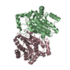
| Noncrystallographic symmetry (NCS) | NCS domain:
NCS domain segments: Component-ID: _ / Beg auth comp-ID: MET / Beg label comp-ID: MET / End auth comp-ID: GLN / End label comp-ID: GLN / Refine code: _ / Auth seq-ID: 1 - 255 / Label seq-ID: 1 - 255
NCS ensembles :
|
-Components
| #1: Protein | Mass: 27350.293 Da / Num. of mol.: 4 Source method: isolated from a genetically manipulated source Source: (gene. exp.) Strain: 168 / Gene: bacC, ywfD, BSU37720, ipa-82d / Plasmid: pET22b / Production host: References: UniProt: P39640, dihydroanticapsin dehydrogenase #2: Chemical | ChemComp-NAI /   Mass: 665.441 Da / Num. of mol.: 4 / Source method: obtained synthetically / Formula: C21H29N7O14P2 Mass: 665.441 Da / Num. of mol.: 4 / Source method: obtained synthetically / Formula: C21H29N7O14P2#3: Water | ChemComp-HOH / |  Mass: 18.015 Da / Num. of mol.: 131 / Source method: isolated from a natural source / Formula: H2O Mass: 18.015 Da / Num. of mol.: 131 / Source method: isolated from a natural source / Formula: H2O |
|---|
-Experimental details
-Experiment
| Experiment | Method: X-RAY DIFFRACTION / Number of used crystals: 1 |
|---|
- Sample preparation
| Crystal | Density Matthews: 2.3 Å3/Da / Density % sol: 46.51 % |
|---|---|
| Crystal grow | Temperature: 291.15 K / Method: microbatch / pH: 6.5 Details: 0.2M Ammonium sulfate, 0.1M BIS-TRIS, pH 6.5, 25% w/v Polyethylene glycol 3350, 10% v/v Glycerol |
-Data collection
| Diffraction | Mean temperature: 100 K |
|---|---|
| Diffraction source | Source: ROTATING ANODE / Type: RIGAKU MICROMAX-007 HF / Wavelength: 1.5418 Å |
| Detector | Type: MAR scanner 345 mm plate / Detector: IMAGE PLATE / Date: Oct 15, 2014 |
| Radiation | Monochromator: VariMax HF / Protocol: SINGLE WAVELENGTH / Monochromatic (M) / Laue (L): M / Scattering type: x-ray |
| Radiation wavelength | Wavelength: 1.5418 Å / Relative weight: 1 |
| Reflection | Resolution: 2.26→38.1 Å / Num. obs: 46073 / % possible obs: 99.8 % / Redundancy: 3.9 % / Biso Wilson estimate: 16.3 Å2 / CC1/2: 0.996 / Rsym value: 0.08 / Net I/σ(I): 12.9 |
| Reflection shell | Resolution: 2.26→2.38 Å / Redundancy: 3.7 % / Rmerge(I) obs: 0.325 / Mean I/σ(I) obs: 3.8 / % possible all: 98.9 |
- Processing
| Software |
| ||||||||||||||||||||||||||||||||||||||||||||||||||||||||||||||||||||||||||||||||||||||||||||||||||||
|---|---|---|---|---|---|---|---|---|---|---|---|---|---|---|---|---|---|---|---|---|---|---|---|---|---|---|---|---|---|---|---|---|---|---|---|---|---|---|---|---|---|---|---|---|---|---|---|---|---|---|---|---|---|---|---|---|---|---|---|---|---|---|---|---|---|---|---|---|---|---|---|---|---|---|---|---|---|---|---|---|---|---|---|---|---|---|---|---|---|---|---|---|---|---|---|---|---|---|---|---|---|
| Refinement | Method to determine structure: MOLECULAR REPLACEMENT / Resolution: 2.26→38.1 Å / Cor.coef. Fo:Fc: 0.927 / Cor.coef. Fo:Fc free: 0.925 / SU B: 6.622 / SU ML: 0.16 / Cross valid method: THROUGHOUT / ESU R: 0.366 / ESU R Free: 0.213
| ||||||||||||||||||||||||||||||||||||||||||||||||||||||||||||||||||||||||||||||||||||||||||||||||||||
| Solvent computation | Ion probe radii: 0.8 Å / Shrinkage radii: 0.8 Å / VDW probe radii: 1.2 Å | ||||||||||||||||||||||||||||||||||||||||||||||||||||||||||||||||||||||||||||||||||||||||||||||||||||
| Displacement parameters | Biso mean: 25.385 Å2
| ||||||||||||||||||||||||||||||||||||||||||||||||||||||||||||||||||||||||||||||||||||||||||||||||||||
| Refinement step | Cycle: 1 / Resolution: 2.26→38.1 Å
| ||||||||||||||||||||||||||||||||||||||||||||||||||||||||||||||||||||||||||||||||||||||||||||||||||||
| Refine LS restraints |
| ||||||||||||||||||||||||||||||||||||||||||||||||||||||||||||||||||||||||||||||||||||||||||||||||||||
| Refine LS restraints NCS | Refine-ID: X-RAY DIFFRACTION / Type: interatomic distance / Weight position: 0.05
| ||||||||||||||||||||||||||||||||||||||||||||||||||||||||||||||||||||||||||||||||||||||||||||||||||||
| LS refinement shell | Resolution: 2.256→2.315 Å / Total num. of bins used: 20
|
