 Movie
Movie Controller

[English] 日本語
 Yorodumi
- PDB-5is1: Crystal structure of the extracellular domain of sensor histidine... -
+ Open data

- Basic information
| Entry | Database: PDB / ID: 5is1 | ||||||
|---|---|---|---|---|---|---|---|
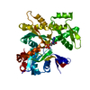
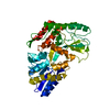
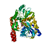
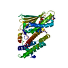
| Title | Crystal structure of the extracellular domain of sensor histidine kinase YycG from Staphylococcus aureus at 2.0 Angstrom resolution | ||||||
 Components Components | Histidine kinase | ||||||
 Keywords Keywords | TRANSFERASE / PAS/PDC domain / sensor histidine kinase / nest motif / membrane protein | ||||||
| Function / homology |  Function and homology information Function and homology informationosmosensory signaling via phosphorelay pathway / phosphorelay response regulator activity / phosphorelay sensor kinase activity / histidine kinase / protein kinase activator activity / ATP binding / metal ion binding / plasma membrane Similarity search - Function | ||||||
| Biological species |   Staphylococcus aureus (bacteria) Staphylococcus aureus (bacteria) | ||||||
| Method |  X-RAY DIFFRACTION / SYNCHROTRON / MOLECULAR REPLACEMENT / Resolution: 1.998 Å X-RAY DIFFRACTION / SYNCHROTRON / MOLECULAR REPLACEMENT / Resolution: 1.998 Å | ||||||
 Authors Authors | Kim, T. / Choi, J. / Lee, S. / Kim, K.K. | ||||||
 Citation Citation | Journal: J.Mol.Biol. / Year: 2016 Title: Structural Studies on the Extracellular Domain of Sensor Histidine Kinase YycG from Staphylococcus aureus and Its Functional Implications Authors: Kim, T. / Choi, J. / Lee, S. / Yeo, K.J. / Cheong, H.K. / Kim, K.K. | ||||||
| History |
|
- Structure visualization
| Structure viewer | Molecule: MolmilJmol/JSmol |
|---|
- Downloads & links
-Download
| PDBx/mmCIF format | 5is1.cif.gz | 74.2 KB | Display | PDBx/mmCIF format |
|---|---|---|---|---|
| PDB format | pdb5is1.ent.gz | 54.4 KB | Display | PDB format |
| PDBx/mmJSON format | 5is1.json.gz | Tree view | PDBx/mmJSON format | |
| Others |  Other downloads Other downloads |
-Validation report
| Arichive directory | https://data.pdbj.org/pub/pdb/validation_reports/is/5is1ftp://data.pdbj.org/pub/pdb/validation_reports/is/5is1 | HTTPS FTP |
|---|
-Related structure data
| Similar structure data |
|---|


-Links
 PDBj
PDBj


- Assembly
| Deposited unit | 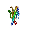
| |||||||||
|---|---|---|---|---|---|---|---|---|---|---|
| 1 | 
| |||||||||
| Unit cell |
| |||||||||
| Components on special symmetry positions |
|
-Components
| #1: Protein | Mass: 17399.508 Da / Num. of mol.: 1 / Fragment: Extracellular domai, UNP residues 33-182 Source method: isolated from a genetically manipulated source Source: (gene. exp.) Staphylococcus aureus (bacteria) / Strain: RN4220Gene: walK, walK_1, AL493_01420, AL498_11610, AL508_14645, AM595_00105, ASU36_11995, AUC48_00105, AUC49_00105, AUC50_00105, BN1321_430109, CH51_00105, EQ80_000105, ER12_000105, ERS092844_02460, ...Gene: walK, walK_1, AL493_01420, AL498_11610, AL508_14645, AM595_00105, ASU36_11995, AUC48_00105, AUC49_00105, AUC50_00105, BN1321_430109, CH51_00105, EQ80_000105, ER12_000105, ERS092844_02460, ERS093009_00906, ERS179246_01836, ERS195423_01765, ERS365775_01678, ERS410449_00558, ERS411009_01438, ERS411017_01103, ERS445051_00023, ERS445052_00659, FE68_05490, NI36_00110, RL02_04070, RT87_00105, SA7112_13655, SASCBU26_00023 Production host: References: UniProt: V5YQR2, UniProt: Q2G2U4*PLUS, histidine kinase |
|---|---|
| #2: Water | ChemComp-HOH /  Mass: 18.015 Da / Num. of mol.: 75 / Source method: isolated from a natural source / Formula: H2O Mass: 18.015 Da / Num. of mol.: 75 / Source method: isolated from a natural source / Formula: H2O |
-Experimental details
-Experiment
| Experiment | Method: X-RAY DIFFRACTION / Number of used crystals: 1 |
|---|
- Sample preparation
| Crystal | Density Matthews: 1.98 Å3/Da / Density % sol: 37.85 % |
|---|---|
| Crystal grow | Temperature: 295 K / Method: microbatch / pH: 6 Details: 40-55% (w/v) PEG 400, 100mM 2-(N-morpholino)ethanesulfonic acid (MES) pH 6.0 |
-Data collection
| Diffraction | Mean temperature: 100 K | |||||||||||||||||||||||||||||||||||||||||||||||||||||||
|---|---|---|---|---|---|---|---|---|---|---|---|---|---|---|---|---|---|---|---|---|---|---|---|---|---|---|---|---|---|---|---|---|---|---|---|---|---|---|---|---|---|---|---|---|---|---|---|---|---|---|---|---|---|---|---|---|
| Diffraction source | Source: SYNCHROTRON / Site: PAL/PLS  / Beamline: 4A / Wavelength: 1 Å / Beamline: 4A / Wavelength: 1 Å | |||||||||||||||||||||||||||||||||||||||||||||||||||||||
| Detector | Type: ADSC QUANTUM 210 / Detector: CCD / Date: Jan 11, 2008 | |||||||||||||||||||||||||||||||||||||||||||||||||||||||
| Radiation | Protocol: SINGLE WAVELENGTH / Monochromatic (M) / Laue (L): M / Scattering type: x-ray | |||||||||||||||||||||||||||||||||||||||||||||||||||||||
| Radiation wavelength | Wavelength: 1 Å / Relative weight: 1 | |||||||||||||||||||||||||||||||||||||||||||||||||||||||
| Reflection | Resolution: 1.998→20 Å / Num. obs: 9779 / % possible obs: 99.4 % / Redundancy: 10.3 % / Biso Wilson estimate: 41.92 Å2 / Rmerge(I) obs: 0.052 / Net I/σ(I): 71.355 | |||||||||||||||||||||||||||||||||||||||||||||||||||||||
| Reflection shell |
|
- Processing
| Software |
| ||||||||||||||||||||||||||||||||||||||||||||||||||||||||
|---|---|---|---|---|---|---|---|---|---|---|---|---|---|---|---|---|---|---|---|---|---|---|---|---|---|---|---|---|---|---|---|---|---|---|---|---|---|---|---|---|---|---|---|---|---|---|---|---|---|---|---|---|---|---|---|---|---|
| Refinement | Method to determine structure: MOLECULAR REPLACEMENT / Resolution: 1.998→19.829 Å / SU ML: 0.14 / Cross valid method: FREE R-VALUE / σ(F): 1.34 / Phase error: 20.87
| ||||||||||||||||||||||||||||||||||||||||||||||||||||||||
| Solvent computation | Shrinkage radii: 0.9 Å / VDW probe radii: 1.11 Å | ||||||||||||||||||||||||||||||||||||||||||||||||||||||||
| Displacement parameters | Biso max: 118.55 Å2 / Biso mean: 54.0375 Å2 / Biso min: 18.17 Å2 | ||||||||||||||||||||||||||||||||||||||||||||||||||||||||
| Refinement step | Cycle: final / Resolution: 1.998→19.829 Å
| ||||||||||||||||||||||||||||||||||||||||||||||||||||||||
| Refine LS restraints |
| ||||||||||||||||||||||||||||||||||||||||||||||||||||||||
| LS refinement shell | Refine-ID: X-RAY DIFFRACTION / Total num. of bins used: 7
|
