| 登録情報 | データベース: PDB / ID: 5ilu
|
|---|
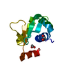
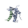
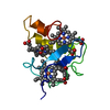
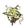
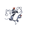
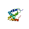
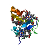
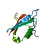
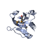
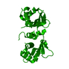
| タイトル | Autoinhibited ETV4 |
|---|
 要素 要素 | ETS translocation variant 4 |
|---|
 キーワード キーワード | DNA BINDING PROTEIN / ETV4 / ETS / transcription factor / autoinhibition transcription / DNA binding |
|---|
| 機能・相同性 |  機能・相同性情報 機能・相同性情報
positive regulation of keratinocyte differentiation / MAPK6/MAPK4 signaling / sequence-specific double-stranded DNA binding / nervous system development / DNA-binding transcription activator activity, RNA polymerase II-specific / DNA-binding transcription factor activity, RNA polymerase II-specific / cell differentiation / RNA polymerase II cis-regulatory region sequence-specific DNA binding / regulation of transcription by RNA polymerase II / chromatin ...positive regulation of keratinocyte differentiation / MAPK6/MAPK4 signaling / sequence-specific double-stranded DNA binding / nervous system development / DNA-binding transcription activator activity, RNA polymerase II-specific / DNA-binding transcription factor activity, RNA polymerase II-specific / cell differentiation / RNA polymerase II cis-regulatory region sequence-specific DNA binding / regulation of transcription by RNA polymerase II / chromatin / DNA-templated transcription / nucleolus / positive regulation of transcription by RNA polymerase II / nucleoplasm / nucleus類似検索 - 分子機能 PEA3-type ETS-domain transcription factor, N-terminal / PEA3 subfamily ETS-domain transcription factor N terminal domain / Ets-domain signature 1. / Ets-domain signature 2. / Ets domain / ETS family / Ets-domain / Ets-domain profile. / erythroblast transformation specific domain / Winged helix-like DNA-binding domain superfamily/Winged helix DNA-binding domain ...PEA3-type ETS-domain transcription factor, N-terminal / PEA3 subfamily ETS-domain transcription factor N terminal domain / Ets-domain signature 1. / Ets-domain signature 2. / Ets domain / ETS family / Ets-domain / Ets-domain profile. / erythroblast transformation specific domain / Winged helix-like DNA-binding domain superfamily/Winged helix DNA-binding domain / Arc Repressor Mutant, subunit A / Winged helix DNA-binding domain superfamily / Winged helix-like DNA-binding domain superfamily / Orthogonal Bundle / Mainly Alpha類似検索 - ドメイン・相同性 |
|---|
| 生物種 |  Homo sapiens (ヒト) Homo sapiens (ヒト) |
|---|
| 手法 |  X線回折 / シンクロトロン / 分子置換 / 解像度: 1.101 Å X線回折 / シンクロトロン / 分子置換 / 解像度: 1.101 Å |
|---|
 データ登録者 データ登録者 | Whitby, F.G. / Currie, S.L. |
|---|
| 資金援助 |  米国, 1件 米国, 1件 | 組織 | 認可番号 | 国 |
|---|
| National Institutes of Health/National Institute of General Medical Sciences (NIH/NIGMS) | R01GM38663 | 米国 |
|
|---|
 引用 引用 | ジャーナル: Nucleic Acids Res. / 年: 2017
タイトル: Structured and disordered regions cooperatively mediate DNA-binding autoinhibition of ETS factors ETV1, ETV4 and ETV5.
著者: Currie, S.L. / Lau, D.K.W. / Doane, J.J. / Whitby, F.G. / Okon, M. / McIntosh, L.P. / Graves, B.J. |
|---|
| 履歴 | | 登録 | 2016年3月4日 | 登録サイト: RCSB / 処理サイト: RCSB |
|---|
| 改定 1.0 | 2017年2月22日 | Provider: repository / タイプ: Initial release |
|---|
| 改定 1.1 | 2017年5月3日 | Group: Database references |
|---|
| 改定 1.2 | 2017年9月20日 | Group: Author supporting evidence / カテゴリ: pdbx_audit_support / Item: _pdbx_audit_support.funding_organization |
|---|
| 改定 1.3 | 2019年12月25日 | Group: Author supporting evidence / カテゴリ: pdbx_audit_support / Item: _pdbx_audit_support.funding_organization |
|---|
| 改定 1.4 | 2024年10月30日 | Group: Data collection / Database references / Structure summary
カテゴリ: chem_comp_atom / chem_comp_bond ...chem_comp_atom / chem_comp_bond / database_2 / pdbx_entry_details / pdbx_modification_feature
Item: _database_2.pdbx_DOI / _database_2.pdbx_database_accession |
|---|
|
|---|
 ムービー
ムービー コントローラー


 データを開く
 基本情報
基本情報 構造の表示
構造の表示 ダウンロードとリンク
 その他のダウンロード
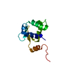

 PDBj
PDBj 集合体
集合体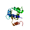

 分子量: 18.015 Da / 分子数: 125 / 由来タイプ: 天然 / 式: H2O
 試料調製
試料調製 解析
解析