 Movie
Movie Controller

[English] 日本語
 Yorodumi
- PDB-5ij6: Crystal structure of Enterococcus faecalis lipoate-protein ligase... -
+ Open data

- Basic information
| Entry | Database: PDB / ID: 5ij6 | ||||||
|---|---|---|---|---|---|---|---|
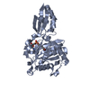
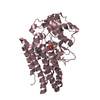
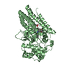
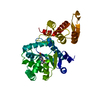
| Title | Crystal structure of Enterococcus faecalis lipoate-protein ligase A (lplA-1) in complex with lipoic acid | ||||||
 Components Components | Lipoate--protein ligase | ||||||
 Keywords Keywords | LIGASE / TRANSFERASE / PROTEIN-SUBSTRATE COMPLEX / LIPOIC ACID | ||||||
| Function / homology |  Function and homology information Function and homology informationlipoyltransferase activity / lipoate-protein ligase / lipoate-protein ligase activity / ATP binding / cytoplasm Similarity search - Function | ||||||
| Biological species |   Enterococcus faecalis (bacteria) Enterococcus faecalis (bacteria) | ||||||
| Method |  X-RAY DIFFRACTION / SYNCHROTRON / MOLECULAR REPLACEMENT / molecular replacement / Resolution: 2 Å X-RAY DIFFRACTION / SYNCHROTRON / MOLECULAR REPLACEMENT / molecular replacement / Resolution: 2 Å | ||||||
 Authors Authors | Hughes, S.J. / Lyle, A.G. / Song, J.H. / Antoshchenko, T. / Park, H. | ||||||
 Citation Citation | Journal: to be published Title: Crystal structure of Enterococcus faecalis lipoate-protein ligase A (lplA-1) in complex with lipoic acid Authors: Hughes, S.J. / Lyle, A.G. / Antoshchenko, T. / Park, H. | ||||||
| History |
|
- Structure visualization
| Structure viewer | Molecule: MolmilJmol/JSmol |
|---|
- Downloads & links
-Download
| PDBx/mmCIF format | 5ij6.cif.gz | 149.4 KB | Display | PDBx/mmCIF format |
|---|---|---|---|---|
| PDB format | pdb5ij6.ent.gz | 116.7 KB | Display | PDB format |
| PDBx/mmJSON format | 5ij6.json.gz | Tree view | PDBx/mmJSON format | |
| Others |  Other downloads Other downloads |
-Validation report
| Summary document | 5ij6_validation.pdf.gz | 449 KB | Display | wwPDB validaton report |
|---|---|---|---|---|
| Full document | 5ij6_full_validation.pdf.gz | 449.7 KB | Display | |
| Data in XML | 5ij6_validation.xml.gz | 14.7 KB | Display | |
| Data in CIF | 5ij6_validation.cif.gz | 21 KB | Display | |
| Arichive directory | https://data.pdbj.org/pub/pdb/validation_reports/ij/5ij6ftp://data.pdbj.org/pub/pdb/validation_reports/ij/5ij6 | HTTPS FTP |
-Related structure data
| Related structure data |  5ibyS  5j3m S: Starting model for refinement |
|---|---|
| Similar structure data |


-Links
 PDBj
PDBj
- Assembly
| Deposited unit | 
| ||||||||
|---|---|---|---|---|---|---|---|---|---|
| 1 |
| ||||||||
| Unit cell |
|
-Components
| #1: Protein | Mass: 40781.395 Da / Num. of mol.: 1 Source method: isolated from a genetically manipulated source Source: (gene. exp.) Enterococcus faecalis (strain ATCC 700802 / V583) (bacteria)Strain: ATCC 700802 / V583 / Gene: lplA-1, EF_0650 / Plasmid: PET28-MHL / Production host: |
|---|---|
| #2: Chemical | ChemComp-LPA /   Mass: 206.326 Da / Num. of mol.: 1 / Source method: obtained synthetically / Formula: C8H14O2S2 Mass: 206.326 Da / Num. of mol.: 1 / Source method: obtained synthetically / Formula: C8H14O2S2 |
| #3: Chemical | ChemComp-SO4 /   Mass: 96.063 Da / Num. of mol.: 1 / Source method: obtained synthetically / Formula: SO4 Mass: 96.063 Da / Num. of mol.: 1 / Source method: obtained synthetically / Formula: SO4 |
| #4: Chemical | ChemComp-CL /   Mass: 35.453 Da / Num. of mol.: 1 / Source method: obtained synthetically / Formula: Cl Mass: 35.453 Da / Num. of mol.: 1 / Source method: obtained synthetically / Formula: Cl |
| #5: Water | ChemComp-HOH /  Mass: 18.015 Da / Num. of mol.: 178 / Source method: isolated from a natural source / Formula: H2O Mass: 18.015 Da / Num. of mol.: 178 / Source method: isolated from a natural source / Formula: H2O |
-Experimental details
-Experiment
| Experiment | Method: X-RAY DIFFRACTION / Number of used crystals: 1 |
|---|
- Sample preparation
| Crystal | Density Matthews: 2.45 Å3/Da / Density % sol: 49.87 % / Mosaicity: 0.33 ° |
|---|---|
| Crystal grow | Temperature: 289 K / Method: vapor diffusion, hanging drop / pH: 8.5 Details: 0.5 UL PROTEIN + 0.5 UL BUFFER (30% PEG 4K, 0.2 M LITHIUM SULFATE, 0.1 M Tris HCl, PH 8.5) |
-Data collection
| Diffraction | Mean temperature: 100 K | ||||||||||||||||||||||||||||||
|---|---|---|---|---|---|---|---|---|---|---|---|---|---|---|---|---|---|---|---|---|---|---|---|---|---|---|---|---|---|---|---|
| Diffraction source | Source: SYNCHROTRON / Site: CLSI  / Beamline: 08ID-1 / Wavelength: 0.97949 Å / Beamline: 08ID-1 / Wavelength: 0.97949 Å | ||||||||||||||||||||||||||||||
| Detector | Type: RAYONIX MX-300 / Detector: CCD / Date: Oct 1, 2015 | ||||||||||||||||||||||||||||||
| Radiation | Monochromator: Si(111) / Protocol: SINGLE WAVELENGTH / Monochromatic (M) / Laue (L): M / Scattering type: x-ray | ||||||||||||||||||||||||||||||
| Radiation wavelength | Wavelength: 0.97949 Å / Relative weight: 1 | ||||||||||||||||||||||||||||||
| Reflection | Resolution: 2→39.44 Å / Num. obs: 27879 / % possible obs: 100 % / Redundancy: 8.6 % / CC1/2: 0.994 / Rmerge(I) obs: 0.145 / Rpim(I) all: 0.052 / Rrim(I) all: 0.154 / Net I/σ(I): 9.1 / Num. measured all: 240100 / Scaling rejects: 44 | ||||||||||||||||||||||||||||||
| Reflection shell | Diffraction-ID: 1
|
-Phasing
| Phasing | Method: molecular replacement | |||||||||
|---|---|---|---|---|---|---|---|---|---|---|
| Phasing MR | Model details: Phaser MODE: MR_AUTO
|
- Processing
| Software |
| |||||||||||||||||||||||||||||||||||||||||||||||||||||||||||||||||||||||||||
|---|---|---|---|---|---|---|---|---|---|---|---|---|---|---|---|---|---|---|---|---|---|---|---|---|---|---|---|---|---|---|---|---|---|---|---|---|---|---|---|---|---|---|---|---|---|---|---|---|---|---|---|---|---|---|---|---|---|---|---|---|---|---|---|---|---|---|---|---|---|---|---|---|---|---|---|---|
| Refinement | Method to determine structure: MOLECULAR REPLACEMENT Starting model: 5IBY Resolution: 2→39.47 Å / Cor.coef. Fo:Fc: 0.956 / Cor.coef. Fo:Fc free: 0.941 / WRfactor Rfree: 0.2121 / WRfactor Rwork: 0.1832 / FOM work R set: 0.8701 / SU B: 7.152 / SU ML: 0.104 / SU R Cruickshank DPI: 0.1571 / SU Rfree: 0.1382 / Cross valid method: THROUGHOUT / σ(F): 0 / ESU R: 0.157 / ESU R Free: 0.138 / Stereochemistry target values: MAXIMUM LIKELIHOOD Details: HYDROGENS HAVE BEEN ADDED IN THE RIDING POSITIONS U VALUES : WITH TLS ADDED
| |||||||||||||||||||||||||||||||||||||||||||||||||||||||||||||||||||||||||||
| Solvent computation | Ion probe radii: 0.7 Å / Shrinkage radii: 0.7 Å / VDW probe radii: 1 Å / Solvent model: MASK | |||||||||||||||||||||||||||||||||||||||||||||||||||||||||||||||||||||||||||
| Displacement parameters | Biso max: 113.98 Å2 / Biso mean: 33.667 Å2 / Biso min: 16.43 Å2
| |||||||||||||||||||||||||||||||||||||||||||||||||||||||||||||||||||||||||||
| Refinement step | Cycle: final / Resolution: 2→39.47 Å
| |||||||||||||||||||||||||||||||||||||||||||||||||||||||||||||||||||||||||||
| Refine LS restraints |
| |||||||||||||||||||||||||||||||||||||||||||||||||||||||||||||||||||||||||||
| LS refinement shell | Resolution: 2→2.108 Å / Rfactor Rfree error: 0 / Total num. of bins used: 10
| |||||||||||||||||||||||||||||||||||||||||||||||||||||||||||||||||||||||||||
| Refinement TLS params. | Method: refined / Refine-ID: X-RAY DIFFRACTION
| |||||||||||||||||||||||||||||||||||||||||||||||||||||||||||||||||||||||||||
| Refinement TLS group |
|
