 Movie
Movie Controller

[English] 日本語
 Yorodumi
- PDB-5ihp: Crystal Structure of Cobyrinic Acid a,c-diamide synthase from Myc... -
+ Open data

- Basic information
| Entry | Database: PDB / ID: 5ihp | ||||||
|---|---|---|---|---|---|---|---|
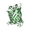
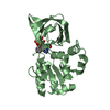
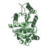
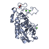
| Title | Crystal Structure of Cobyrinic Acid a,c-diamide synthase from Mycobacterium smegmatis with bound ADP and Magnesium | ||||||
 Components Components | Cobyrinic Acid a,c-diamide synthase | ||||||
 Keywords Keywords | LIGASE / SSGCID / COBYRINIC ACID A / C-DIAMIDE SYNTHASE / Structural Genomics / Seattle Structural Genomics Center for Infectious Disease | ||||||
| Function / homology |  Function and homology information Function and homology information | ||||||
| Biological species |  Mycobacterium smegmatis (bacteria) Mycobacterium smegmatis (bacteria) | ||||||
| Method |  X-RAY DIFFRACTION / SYNCHROTRON / MOLECULAR REPLACEMENT / Resolution: 1.85 Å X-RAY DIFFRACTION / SYNCHROTRON / MOLECULAR REPLACEMENT / Resolution: 1.85 Å | ||||||
 Authors Authors | Seattle Structural Genomics Center for Infectious Disease (SSGCID) | ||||||
 Citation Citation | Journal: TO BE PUBLISHED Title: Crystal Structure of Cobyrinic Acid a,c-diamide synthase from Mycobacterium smegmatis with bound ATP analog and Magnesium Authors: Davies, D.R. / Horanyi, P.S. / Abendroth, J.A. / SSGCID | ||||||
| History |
|
- Structure visualization
| Structure viewer | Molecule: MolmilJmol/JSmol |
|---|
- Downloads & links
-Download
| PDBx/mmCIF format | 5ihp.cif.gz | 202.1 KB | Display | PDBx/mmCIF format |
|---|---|---|---|---|
| PDB format | pdb5ihp.ent.gz | 159.5 KB | Display | PDB format |
| PDBx/mmJSON format | 5ihp.json.gz | Tree view | PDBx/mmJSON format | |
| Others |  Other downloads Other downloads |
-Validation report
| Arichive directory | https://data.pdbj.org/pub/pdb/validation_reports/ih/5ihpftp://data.pdbj.org/pub/pdb/validation_reports/ih/5ihp | HTTPS FTP |
|---|
-Related structure data
| Related structure data |  5if9C  4pfsS S: Starting model for refinement C: citing same article ( |
|---|---|
| Similar structure data | |
| Other databases |


-Links
 PDBj
PDBj- Assembly
| Deposited unit | 
| ||||||||
|---|---|---|---|---|---|---|---|---|---|
| 1 | 
| ||||||||
| 2 | 
| ||||||||
| Unit cell |
| ||||||||
| Components on special symmetry positions |
|
-Components
| #1: Protein | Mass: 29410.924 Da / Num. of mol.: 2 Source method: isolated from a genetically manipulated source Source: (gene. exp.) Mycobacterium smegmatis (strain ATCC 700084 / mc(2)155) (bacteria)Strain: ATCC 700084 / mc(2)155 / Gene: MSMEG_1927 / Production host: #2: Chemical |   Mass: 427.201 Da / Num. of mol.: 2 / Source method: obtained synthetically / Formula: C10H15N5O10P2 / Comment: ADP, energy-carrying molecule*YM Mass: 427.201 Da / Num. of mol.: 2 / Source method: obtained synthetically / Formula: C10H15N5O10P2 / Comment: ADP, energy-carrying molecule*YM#3: Chemical |   Mass: 24.305 Da / Num. of mol.: 2 / Source method: obtained synthetically / Formula: Mg Mass: 24.305 Da / Num. of mol.: 2 / Source method: obtained synthetically / Formula: Mg#4: Water | ChemComp-HOH / |  Mass: 18.015 Da / Num. of mol.: 277 / Source method: isolated from a natural source / Formula: H2O Mass: 18.015 Da / Num. of mol.: 277 / Source method: isolated from a natural source / Formula: H2O |
|---|
-Experimental details
-Experiment
| Experiment | Method: X-RAY DIFFRACTION / Number of used crystals: 1 |
|---|
- Sample preparation
| Crystal | Density Matthews: 2.01 Å3/Da / Density % sol: 38.92 % |
|---|---|
| Crystal grow | Temperature: 289 K / Method: vapor diffusion, sitting drop / pH: 8 Details: EBS INTERNAL TRACKING NUMBER 500 MM NACL, 2 MM DTT, 0.025% SODIUM AZIDE, 5% GLYCEROL, 0.4 UL X 0.4 UL DROP WITH JCSG SCREEN CONDITION F3: 20% (V/V) MPD, 100 MM TRIS PH 8.0; 3 DAY SOAK WITH 5 ...Details: EBS INTERNAL TRACKING NUMBER 500 MM NACL, 2 MM DTT, 0.025% SODIUM AZIDE, 5% GLYCEROL, 0.4 UL X 0.4 UL DROP WITH JCSG SCREEN CONDITION F3: 20% (V/V) MPD, 100 MM TRIS PH 8.0; 3 DAY SOAK WITH 5 MM ADP + 5 MM MGCL2; 20% ETHYLENE GLYCOL CRYOPROTECTANT PH range: 8 |
-Data collection
| Diffraction | Mean temperature: 100 K |
|---|---|
| Diffraction source | Source: SYNCHROTRON / Site: APS  / Beamline: 21-ID-F / Wavelength: 0.97872 Å / Beamline: 21-ID-F / Wavelength: 0.97872 Å |
| Detector | Type: MARMOSAIC 225 mm CCD / Detector: CCD / Date: Jul 30, 2015 |
| Radiation | Protocol: SINGLE WAVELENGTH / Monochromatic (M) / Laue (L): M / Scattering type: x-ray |
| Radiation wavelength | Wavelength: 0.97872 Å / Relative weight: 1 |
| Reflection | Resolution: 1.85→42 Å / Num. obs: 41651 / % possible obs: 100 % / Observed criterion σ(I): -3 / Redundancy: 12.07 % / Biso Wilson estimate: 30.38 Å2 / Rmerge(I) obs: 0.06 / Net I/σ(I): 29.11 |
| Reflection shell | Resolution: 1.85→1.9 Å / Rmerge(I) obs: 0.542 / Mean I/σ(I) obs: 4.9 / % possible all: 100 |
- Processing
| Software |
| ||||||||||||||||
|---|---|---|---|---|---|---|---|---|---|---|---|---|---|---|---|---|---|
| Refinement | Method to determine structure: MOLECULAR REPLACEMENT Starting model: 4PFS Resolution: 1.85→41.63 Å / Cross valid method: FREE R-VALUE
| ||||||||||||||||
| Displacement parameters | Biso mean: 28.94 Å2 | ||||||||||||||||
| Refinement step | Cycle: LAST / Resolution: 1.85→41.63 Å
|
