| Entry | Database: PDB / ID: 5ihb
|
|---|
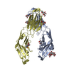
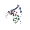
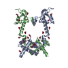
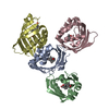
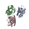
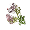
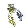
| Title | Structure of the immune receptor CD33 |
|---|
 Components Components | Myeloid cell surface antigen CD33 |
|---|
 Keywords Keywords | IMMUNE SYSTEM / Immune receptor / Siglec / Ig-like / sialic-acid binding |
|---|
| Function / homology |  Function and homology information Function and homology information
immune response-inhibiting signal transduction / protein tyrosine phosphatase activator activity / negative regulation of monocyte activation / sialic acid binding / Fc-gamma receptor signaling pathway / negative regulation of interleukin-8 production / negative regulation of interleukin-1 beta production / tertiary granule membrane / negative regulation of tumor necrosis factor production / specific granule membrane ...immune response-inhibiting signal transduction / protein tyrosine phosphatase activator activity / negative regulation of monocyte activation / sialic acid binding / Fc-gamma receptor signaling pathway / negative regulation of interleukin-8 production / negative regulation of interleukin-1 beta production / tertiary granule membrane / negative regulation of tumor necrosis factor production / specific granule membrane / positive regulation of protein secretion / cell-cell adhesion / Immunoregulatory interactions between a Lymphoid and a non-Lymphoid cell / cell-cell signaling / peroxisome / carbohydrate binding / signaling receptor activity / protein phosphatase binding / cell adhesion / negative regulation of cell population proliferation / external side of plasma membrane / Neutrophil degranulation / Golgi apparatus / cell surface / signal transduction / plasma membraneSimilarity search - Function : / Immunoglobulin / Immunoglobulin domain / Immunoglobulin V-set domain / Immunoglobulin V-set domain / Immunoglobulin subtype / Immunoglobulin / Ig-like domain profile. / Immunoglobulin-like domain / Immunoglobulin-like domain superfamily ...: / Immunoglobulin / Immunoglobulin domain / Immunoglobulin V-set domain / Immunoglobulin V-set domain / Immunoglobulin subtype / Immunoglobulin / Ig-like domain profile. / Immunoglobulin-like domain / Immunoglobulin-like domain superfamily / Immunoglobulin-like fold / Immunoglobulins / Immunoglobulin-like / Sandwich / Mainly BetaSimilarity search - Domain/homology |
|---|
| Biological species |  Homo sapiens (human) Homo sapiens (human) |
|---|
| Method |  X-RAY DIFFRACTION / SYNCHROTRON / MOLECULAR REPLACEMENT / Resolution: 2.24 Å X-RAY DIFFRACTION / SYNCHROTRON / MOLECULAR REPLACEMENT / Resolution: 2.24 Å |
|---|
 Authors Authors | Dodd, R.B. |
|---|
| Funding support |  United Kingdom, 1items United Kingdom, 1items | Organization | Grant number | Country |
|---|
| Wellcome Trust | RG47376 | United Kingdom |
|
|---|
 Citation Citation | Journal: To Be Published
Title: Structure of ligand bound CD33 receptor associated with Alzheimer's disease
Authors: Dodd, R.B. / Meadows, W. / Qamar, S. / Johnson, C.M. / Kronenberg-Versteeg, D. / St George-Hyslop, P. |
|---|
| History | | Deposition | Feb 29, 2016 | Deposition site: RCSB / Processing site: PDBE |
|---|
| Revision 1.0 | Mar 29, 2017 | Provider: repository / Type: Initial release |
|---|
| Revision 1.1 | Oct 2, 2019 | Group: Data collection / Derived calculations / Category: pdbx_struct_assembly_gen / pdbx_struct_oper_list |
|---|
| Revision 1.2 | Jul 29, 2020 | Group: Data collection / Derived calculations / Structure summary
Category: chem_comp / entity ...chem_comp / entity / pdbx_chem_comp_identifier / pdbx_entity_nonpoly / struct_conn / struct_site / struct_site_gen
Item: _chem_comp.name / _chem_comp.type ..._chem_comp.name / _chem_comp.type / _entity.pdbx_description / _pdbx_entity_nonpoly.name / _struct_conn.pdbx_role
Description: Carbohydrate remediation / Provider: repository / Type: Remediation |
|---|
| Revision 1.3 | Jan 10, 2024 | Group: Data collection / Database references ...Data collection / Database references / Refinement description / Structure summary
Category: chem_comp / chem_comp_atom ...chem_comp / chem_comp_atom / chem_comp_bond / database_2 / pdbx_initial_refinement_model
Item: _chem_comp.pdbx_synonyms / _database_2.pdbx_DOI / _database_2.pdbx_database_accession |
|---|
| Revision 1.4 | Nov 13, 2024 | Group: Structure summary / Category: pdbx_entry_details / pdbx_modification_feature |
|---|
|
|---|
 Movie
Movie Controller


 Open data
 Basic information
 Structure visualization
 Downloads & links
 Other downloads


 PDBj
PDBj


 Assembly


 Type: D-saccharide, beta linking / Mass: 221.208 Da / Num. of mol.: 12 / Source method: isolated from a natural source / Formula: C8H15NO6
 Mass: 18.015 Da / Num. of mol.: 232 / Source method: isolated from a natural source / Formula: H2O
 Sample preparation
 Processing