 Movie
Movie Controller


+ Open data

- Basic information
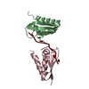
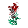
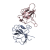
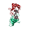
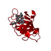
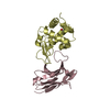
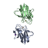
| Entry | Database: PDB / ID: 5ibw | ||||||
|---|---|---|---|---|---|---|---|
| Title | Complex of MlcC bound to the tandem IQ motif of MyoC | ||||||
 Components Components |
| ||||||
 Keywords Keywords | MOTOR PROTEIN / Myosin IQ motif Complex | ||||||
| Function / homology |  Function and homology information Function and homology informationphagocytic cup lip / actin wave / macropinocytic cup cytoskeleton / actin cortical patch localization / pinocytosis / myosin I binding / macropinocytic cup / myosin light chain binding / mitotic spindle microtubule / filopodium assembly ...phagocytic cup lip / actin wave / macropinocytic cup cytoskeleton / actin cortical patch localization / pinocytosis / myosin I binding / macropinocytic cup / myosin light chain binding / mitotic spindle microtubule / filopodium assembly / myosin complex / microfilament motor activity / cell leading edge / phagocytosis / enzyme regulator activity / mitotic spindle organization / actin filament organization / cell motility / phospholipid binding / microtubule cytoskeleton organization / chemotaxis / endocytosis / actin filament binding / lamellipodium / actin cytoskeleton / actin cytoskeleton organization / microtubule binding / calcium ion binding / ATP binding / plasma membrane / cytoplasm / cytosol Similarity search - Function | ||||||
| Biological species |  | ||||||
| Method |  X-RAY DIFFRACTION / SYNCHROTRON / MOLECULAR REPLACEMENT / Resolution: 1.9 Å X-RAY DIFFRACTION / SYNCHROTRON / MOLECULAR REPLACEMENT / Resolution: 1.9 Å | ||||||
 Authors Authors | Langelaan, D.N. / Smith, S.P. | ||||||
| Funding support |  Canada, 1items Canada, 1items
| ||||||
 Citation Citation | Journal: J.Biol.Chem. / Year: 2016 Title: Structure of the Single-lobe Myosin Light Chain C in Complex with the Light Chain-binding Domains of Myosin-1C Provides Insights into Divergent IQ Motif Recognition. Authors: Langelaan, D.N. / Liburd, J. / Yang, Y. / Miller, E. / Chitayat, S. / Crawley, S.W. / Cote, G.P. / Smith, S.P. | ||||||
| History |
|
- Structure visualization
| Structure viewer | Molecule: MolmilJmol/JSmol |
|---|
- Downloads & links
-Download
| PDBx/mmCIF format | 5ibw.cif.gz | 59.1 KB | Display | PDBx/mmCIF format |
|---|---|---|---|---|
| PDB format | pdb5ibw.ent.gz | 41.6 KB | Display | PDB format |
| PDBx/mmJSON format | 5ibw.json.gz | Tree view | PDBx/mmJSON format | |
| Others |  Other downloads Other downloads |
-Validation report
| Arichive directory | https://data.pdbj.org/pub/pdb/validation_reports/ib/5ibwftp://data.pdbj.org/pub/pdb/validation_reports/ib/5ibw | HTTPS FTP |
|---|
-Related structure data
| Related structure data | 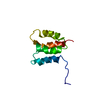 2m8uC 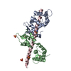 2ix7S S: Starting model for refinement C: citing same article ( |
|---|---|
| Similar structure data |


-Links
 PDBj
PDBj


- Assembly
| Deposited unit | 
| ||||||||
|---|---|---|---|---|---|---|---|---|---|
| 1 |
| ||||||||
| Unit cell |
|
-Components
| #1: Protein | Mass: 8917.953 Da / Num. of mol.: 2 Source method: isolated from a genetically manipulated source Source: (gene. exp.)  #2: Protein/peptide | | Mass: 5464.219 Da / Num. of mol.: 1 / Fragment: UNP residues 699-739 Source method: isolated from a genetically manipulated source Source: (gene. exp.) #3: Chemical | ChemComp-NA / |   Mass: 22.990 Da / Num. of mol.: 1 / Source method: obtained synthetically / Formula: Na Mass: 22.990 Da / Num. of mol.: 1 / Source method: obtained synthetically / Formula: Na#4: Water | ChemComp-HOH / |  Mass: 18.015 Da / Num. of mol.: 238 / Source method: isolated from a natural source / Formula: H2O Mass: 18.015 Da / Num. of mol.: 238 / Source method: isolated from a natural source / Formula: H2O |
|---|
-Experimental details
-Experiment
| Experiment | Method: X-RAY DIFFRACTION / Number of used crystals: 1 |
|---|
- Sample preparation
| Crystal | Density Matthews: 1.96 Å3/Da / Density % sol: 37.13 % / Description: Small triangular prisms |
|---|---|
| Crystal grow | Temperature: 298 K / Method: vapor diffusion, hanging drop / pH: 5.6 / Details: 0.7M NaH2PO4/K2HPO4 pH 5.6 |
-Data collection
| Diffraction | Mean temperature: 100 K |
|---|---|
| Diffraction source | Source: SYNCHROTRON / Site: CLSI / Beamline: 08ID-1 / Wavelength: 1.7712 Å |
| Detector | Type: MARMOSAIC 300 mm CCD / Detector: CCD / Date: Feb 12, 2016 |
| Radiation | Protocol: SINGLE WAVELENGTH / Monochromatic (M) / Laue (L): M / Scattering type: x-ray |
| Radiation wavelength | Wavelength: 1.7712 Å / Relative weight: 1 |
| Reflection | Resolution: 1.9→28.2 Å / Num. obs: 12613 / % possible obs: 90.9 % / Redundancy: 3.8 % / Rmerge(I) obs: 0.043 / Net I/σ(I): 26.9 |
| Reflection shell | Resolution: 1.9→1.97 Å |
- Processing
| Software |
| ||||||||||||||||||
|---|---|---|---|---|---|---|---|---|---|---|---|---|---|---|---|---|---|---|---|
| Refinement | Method to determine structure: MOLECULAR REPLACEMENT Starting model: 2IX7 Resolution: 1.9→28.2 Å / Cross valid method: FREE R-VALUE
| ||||||||||||||||||
| Displacement parameters | Biso max: 50.42 Å2 / Biso mean: 16.7145 Å2 / Biso min: 4.77 Å2 | ||||||||||||||||||
| Refinement step | Cycle: LAST / Resolution: 1.9→28.2 Å
| ||||||||||||||||||
| LS refinement shell | Resolution: 1.9→2.05 Å / Rfactor Rfree: 0.19 / Rfactor Rwork: 0.12 |
