| 登録情報 | データベース: PDB / ID: 5i4h
|
|---|
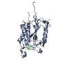
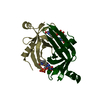
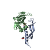
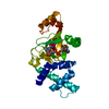
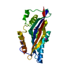
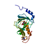
| タイトル | Caught in the Act: The Crystal Structure of cleaved Cathepsin L bound to the active site of Cathepsin L |
|---|
 要素 要素 | (Cathepsin L1) x 2 |
|---|
 キーワード キーワード | HYDROLASE / cathepsin / cysteine cathepsin / substrate / interaction |
|---|
| 機能・相同性 |  機能・相同性情報 機能・相同性情報
enkephalin processing / cathepsin L / CD4-positive, alpha-beta T cell lineage commitment / macrophage apoptotic process / chromaffin granule / elastin catabolic process / antigen processing and presentation of peptide antigen / RUNX1 regulates transcription of genes involved in differentiation of keratinocytes / endolysosome lumen / cellular response to thyroid hormone stimulus ...enkephalin processing / cathepsin L / CD4-positive, alpha-beta T cell lineage commitment / macrophage apoptotic process / chromaffin granule / elastin catabolic process / antigen processing and presentation of peptide antigen / RUNX1 regulates transcription of genes involved in differentiation of keratinocytes / endolysosome lumen / cellular response to thyroid hormone stimulus / Trafficking and processing of endosomal TLR / proteoglycan binding / zymogen activation / Assembly of collagen fibrils and other multimeric structures / antigen processing and presentation / protein autoprocessing / Collagen degradation / collagen catabolic process / fibronectin binding / serpin family protein binding / collagen binding / Attachment and Entry / Degradation of the extracellular matrix / receptor-mediated endocytosis of virus by host cell / multivesicular body / endocytic vesicle lumen / MHC class II antigen presentation / cysteine-type peptidase activity / lysosomal lumen / proteolysis involved in protein catabolic process / Endosomal/Vacuolar pathway / antigen processing and presentation of exogenous peptide antigen via MHC class II / : / histone binding / adaptive immune response / Attachment and Entry / lysosome / apical plasma membrane / fusion of virus membrane with host plasma membrane / cysteine-type endopeptidase activity / intracellular membrane-bounded organelle / fusion of virus membrane with host endosome membrane / symbiont entry into host cell / Golgi apparatus / proteolysis / extracellular space / extracellular exosome / extracellular region / nucleus / plasma membrane類似検索 - 分子機能 Cathepsin propeptide inhibitor domain (I29) / Cathepsin propeptide inhibitor domain (I29) / Cathepsin propeptide inhibitor domain (I29) / Papain-like cysteine endopeptidase / Cysteine peptidase, asparagine active site / Eukaryotic thiol (cysteine) proteases asparagine active site. / Cysteine peptidase, histidine active site / Eukaryotic thiol (cysteine) proteases histidine active site. / : / Peptidase C1A, papain C-terminal ...Cathepsin propeptide inhibitor domain (I29) / Cathepsin propeptide inhibitor domain (I29) / Cathepsin propeptide inhibitor domain (I29) / Papain-like cysteine endopeptidase / Cysteine peptidase, asparagine active site / Eukaryotic thiol (cysteine) proteases asparagine active site. / Cysteine peptidase, histidine active site / Eukaryotic thiol (cysteine) proteases histidine active site. / : / Peptidase C1A, papain C-terminal / Papain family cysteine protease / Papain family cysteine protease / Cysteine peptidase, cysteine active site / Eukaryotic thiol (cysteine) proteases cysteine active site. / Papain-like cysteine peptidase superfamily類似検索 - ドメイン・相同性 |
|---|
| 生物種 |  Homo sapiens (ヒト) Homo sapiens (ヒト) |
|---|
| 手法 |  X線回折 / シンクロトロン / 分子置換 / 解像度: 1.42 Å X線回折 / シンクロトロン / 分子置換 / 解像度: 1.42 Å |
|---|
 データ登録者 データ登録者 | Sosnowski, P. / Turk, D. |
|---|
 引用 引用 | ジャーナル: Febs Lett. / 年: 2016
タイトル: Caught in the act: the crystal structure of cleaved cathepsin L bound to the active site of Cathepsin L.
著者: Sosnowski, P. / Turk, D. |
|---|
| 履歴 | | 登録 | 2016年2月12日 | 登録サイト: RCSB / 処理サイト: PDBE |
|---|
| 改定 1.0 | 2016年4月13日 | Provider: repository / タイプ: Initial release |
|---|
| 改定 1.1 | 2024年1月10日 | Group: Data collection / Database references / Refinement description
カテゴリ: chem_comp_atom / chem_comp_bond ...chem_comp_atom / chem_comp_bond / database_2 / pdbx_initial_refinement_model
Item: _database_2.pdbx_DOI / _database_2.pdbx_database_accession |
|---|
| 改定 1.2 | 2024年11月13日 | Group: Structure summary
カテゴリ: pdbx_entry_details / pdbx_modification_feature |
|---|
|
|---|
 ムービー
ムービー コントローラー


 データを開く
 基本情報
基本情報 構造の表示
構造の表示 ダウンロードとリンク
 その他のダウンロード


 PDBj
PDBj


 集合体
集合体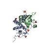
 Komagataella pastoris GS115 (菌類) / 参照: UniProt: P07711, cathepsin L
 分子量: 96.063 Da / 分子数: 4 / 由来タイプ: 組換発現 / 式: SO4
 分子量: 92.094 Da / 分子数: 2 / 由来タイプ: 合成 / 式: C3H8O3
 分子量: 18.015 Da / 分子数: 246 / 由来タイプ: 天然 / 式: H2O
 試料調製
試料調製 / ビームライン: 14.1 / 波長: 1 Å
 解析
解析