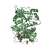
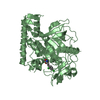
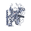
- PDB-5i35: Structure of the Human mitochondrial kinase COQ8A R611K with AMPP... -
+
Open data
ID or keywords:
Loading...
-
Basic information
Entry
Database: PDB / ID: 5i35
Title
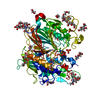
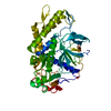
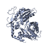
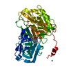
Structure of the Human mitochondrial kinase COQ8A R611K with AMPPNP (Cerebellar Ataxia and Ubiquinone Deficiency Through Loss of Unorthodox Kinase Activity)
Mass: 18.015 Da / Num. of mol.: 26 / Source method: isolated from a natural source / Formula: H2O
Has protein modification
Y
-
Experimental details
-
Experiment
Experiment
Method: X-RAY DIFFRACTION / Number of used crystals: 1
-
Sample preparation
Crystal
Density Matthews: 2.44 Å3/Da / Density % sol: 49.61 %
Crystal grow
Temperature: 293 K / Method: vapor diffusion, hanging drop / pH: 7.5 Details: One part of ADCK3 R611K, 0.2 mM, AMPPNP 2 mM, MgCl2, 4mM, mixed with an equal volume of sodium polyacrylate 5100, 26%; magnesium chloride, 20 mM; HEPES, 100 mM, pH 7.5
Protocol: SINGLE WAVELENGTH / Monochromatic (M) / Laue (L): M / Scattering type: x-ray
Radiation wavelength
Wavelength: 0.96862 Å / Relative weight: 1
Reflection
Resolution: 2.3→50 Å / Num. obs: 19840 / % possible obs: 100 % / Redundancy: 7.37 % / CC1/2: 0.998 / Rmerge(I) obs: 0.087 / Net I/σ(I): 12.5
Reflection shell
Resolution: 2.3→2.36 Å / Redundancy: 7.61 % / Rmerge(I) obs: 1.232 / Mean I/σ(I) obs: 1.65 / % possible all: 100
-
Processing
Software
Name
Version
Classification
REFMAC
5.8.0135
refinement
XDS
datareduction
XSCALE
datascaling
PHASER
phasing
Refinement
Method to determine structure: SAD / Resolution: 2.3→44.67 Å / Cor.coef. Fo:Fc: 0.966 / Cor.coef. Fo:Fc free: 0.922 / SU B: 18.075 / SU ML: 0.194 / Cross valid method: THROUGHOUT / ESU R: 0.325 / ESU R Free: 0.255 / Stereochemistry target values: MAXIMUM LIKELIHOOD / Details: HYDROGENS HAVE BEEN ADDED IN THE RIDING POSITIONS
Rfactor
Num. reflection
% reflection
Selection details
Rfree
0.25887
1556
7.8 %
RANDOM
Rwork
0.18346
-
-
-
obs
0.18952
18275
99.96 %
-
Solvent computation
Ion probe radii: 0.8 Å / Shrinkage radii: 0.8 Å / VDW probe radii: 1.2 Å / Solvent model: BABINET MODEL WITH MASK
 Movie
Movie Controller

 Yorodumi
 Open data
 Basic information
 Components
 Keywords
 Function and homology information
 Homo sapiens (human)
 X-RAY DIFFRACTION /
  Authors
 United States, 2items
  Citation
 Structure visualization
 Downloads & links
 Other downloads
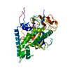


 PDBj
PDBj


 Assembly
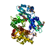


 Mass: 506.196 Da / Num. of mol.: 1 / Source method: obtained synthetically / Formula: C10H17N6O12P3 / Comment: AMP-PNP, energy-carrying molecule analogue*YM
 Mass: 18.015 Da / Num. of mol.: 26 / Source method: isolated from a natural source / Formula: H2O
 Sample preparation
 Processing