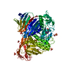
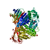
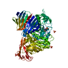
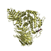
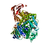
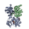
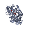
Entry Database : PDB / ID : 5i23Title Crystal Structure of Agd31B, alpha-transglucosylase in Glycoside Hydrolase Family 31, in complex with Cyclophellitol Aziridine probe CF022 Oligosaccharide 4-alpha-D-glucosyltransferase Keywords / / / / Function / homology Function Domain/homology Component
/ / / / / / / / / / / / / / / / / / / / / / / / / / / / / / / / Biological species Cellvibrio japonicus (bacteria)Method / / / Resolution : 1.95 Å Authors Wu, L. / Davies, G.J. Funding support Organization Grant number Country European Research Council ERC-2012-AdG-322942
Journal : Acs Cent.Sci. / Year : 2016Title : Detection of Active Mammalian GH31 alpha-Glucosidases in Health and Disease Using In-Class, Broad-Spectrum Activity-Based Probes.Authors : Jiang, J. / Kuo, C.L. / Wu, L. / Franke, C. / Kallemeijn, W.W. / Florea, B.I. / van Meel, E. / van der Marel, G.A. / Codee, J.D. / Boot, R.G. / Davies, G.J. / Overkleeft, H.S. / Aerts, J.M. History Deposition Feb 8, 2016 Deposition site / Processing site Revision 1.0 May 4, 2016 Provider / Type Revision 1.1 Jun 22, 2016 Group Revision 1.2 Jan 10, 2024 Group / Database references / Refinement descriptionCategory chem_comp_atom / chem_comp_bond ... chem_comp_atom / chem_comp_bond / database_2 / pdbx_initial_refinement_model Item / _database_2.pdbx_database_accessionRevision 1.3 Nov 20, 2024 Group / Category / pdbx_modification_feature
Show all Show less
 Movie
Movie Controller

 Yorodumi
 Open data
 Basic information
 Components
 Keywords
 Function and homology information
 Cellvibrio japonicus (bacteria)
 X-RAY DIFFRACTION /
  Authors
 United Kingdom, 1items
  Citation
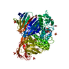
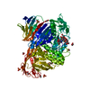
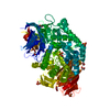
 Structure visualization
 Downloads & links
 Other downloads
 PDBj
PDBj


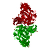
 Assembly


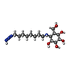

 Mass: 194.226 Da / Num. of mol.: 1 / Source method: obtained synthetically / Formula: C8H18O5 / Comment: precipitant*YM
 Mass: 96.063 Da / Num. of mol.: 6 / Source method: obtained synthetically / Formula: SO4
 Mass: 62.068 Da / Num. of mol.: 2 / Source method: obtained synthetically / Formula: C2H6O2
 Mass: 347.430 Da / Num. of mol.: 1 / Source method: obtained synthetically / Formula: C15H31N4O5
 Sample preparation
 Processing