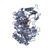
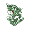
Deposited unit
A: L-amino acid deaminase
B: L-amino acid deaminase
C: L-amino acid deaminase
D: L-amino acid deaminase
E: L-amino acid deaminase
F: L-amino acid deaminase
hetero molecules Summary Component details
Theoretical mass Number of molelcules Total (without water) 309,442 27 Polymers 300,460 6 Non-polymers 8,981 21 Water 1,910 106
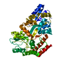
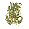
1
A: L-amino acid deaminase
hetero molecules Summary Component details Symmetry operations Calculated values
Theoretical mass Number of molelcules Total (without water) 51,716 5 Polymers 50,077 1 Non-polymers 1,639 4 Water 18 1
Type Name Symmetry operation Number identity operation 1_555 x,y,z 1
Buried area 1420 Å2 ΔGint -9 kcal/mol Surface area 18230 Å2 Method
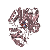
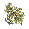
2
B: L-amino acid deaminase
hetero molecules Summary Component details Symmetry operations Calculated values
Theoretical mass Number of molelcules Total (without water) 52,285 7 Polymers 50,077 1 Non-polymers 2,208 6 Water 18 1
Type Name Symmetry operation Number identity operation 1_555 x,y,z 1
Buried area 1440 Å2 ΔGint -9 kcal/mol Surface area 18120 Å2 Method
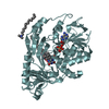
3
C: L-amino acid deaminase
hetero molecules Summary Component details Symmetry operations Calculated values
Theoretical mass Number of molelcules Total (without water) 51,147 3 Polymers 50,077 1 Non-polymers 1,070 2 Water 18 1
Type Name Symmetry operation Number identity operation 1_555 x,y,z 1
Buried area 1460 Å2 ΔGint -9 kcal/mol Surface area 17950 Å2 Method
4
D: L-amino acid deaminase
hetero molecules Summary Component details Symmetry operations Calculated values
Theoretical mass Number of molelcules Total (without water) 51,716 5 Polymers 50,077 1 Non-polymers 1,639 4 Water 18 1
Type Name Symmetry operation Number identity operation 1_555 x,y,z 1
Buried area 1440 Å2 ΔGint -9 kcal/mol Surface area 18030 Å2 Method
5
E: L-amino acid deaminase
hetero molecules Summary Component details Symmetry operations Calculated values
Theoretical mass Number of molelcules Total (without water) 51,147 3 Polymers 50,077 1 Non-polymers 1,070 2 Water 18 1
Type Name Symmetry operation Number identity operation 1_555 x,y,z 1
Buried area 1450 Å2 ΔGint -9 kcal/mol Surface area 18040 Å2 Method
6
F: L-amino acid deaminase
hetero molecules Summary Component details Symmetry operations Calculated values
Theoretical mass Number of molelcules Total (without water) 51,431 4 Polymers 50,077 1 Non-polymers 1,355 3 Water 18 1
Type Name Symmetry operation Number identity operation 1_555 x,y,z 1
Buried area 1440 Å2 ΔGint -9 kcal/mol Surface area 18120 Å2 Method
Unit cell Length a, b, c (Å) 100.314, 104.575, 105.419 Angle α, β, γ (deg.) 64.52, 73.06, 61.17 Int Tables number 1 Space group name H-M P1
 Movie
Movie Controller


 Open data
 Basic information
 Components
 Keywords
 Function and homology information
 Proteus vulgaris (bacteria)
 X-RAY DIFFRACTION /
  Authors
 Citation
 Structure visualization
 Downloads & links
 Other downloads


 PDBj
PDBj Assembly


 Mass: 785.550 Da / Num. of mol.: 6 / Source method: obtained synthetically / Formula: C27H33N9O15P2 / Comment: FAD*YM
 Mass: 284.543 Da / Num. of mol.: 15 / Source method: obtained synthetically / Formula: C19H42N
 Mass: 18.015 Da / Num. of mol.: 106 / Source method: isolated from a natural source / Formula: H2O
 Sample preparation
 / Beamline: 3W1A / Wavelength: 1 Å
 Processing