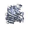
[English] 日本語
Yorodumi
- PDB-5hsf: 1.52 Angstrom Crystal Structure of Fc fragment of Human IgG1. -

+
Open data


ID or keywords:

Loading...

-
Basic information

Entry
Database: PDB / ID: 5hsf
Title1.52 Angstrom Crystal Structure of Fc fragment of Human IgG1.
ComponentsIg gamma-1 chain C region
KeywordsIMMUNE SYSTEM / Fc fragment / IgG1 / Structural Genomics / Center for Structural Genomics of Infectious Diseases / CSGID
Function / homology
Function and homology information


Fc-gamma receptor I complex binding / complement-dependent cytotoxicity / IgG immunoglobulin complex / antibody-dependent cellular cytotoxicity / Classical antibody-mediated complement activation / Initial triggering of complement / FCGR activation / Role of phospholipids in phagocytosis / immunoglobulin complex, circulating / immunoglobulin receptor binding ...Fc-gamma receptor I complex binding / complement-dependent cytotoxicity / IgG immunoglobulin complex / antibody-dependent cellular cytotoxicity / Classical antibody-mediated complement activation / Initial triggering of complement / FCGR activation / Role of phospholipids in phagocytosis / immunoglobulin complex, circulating / immunoglobulin receptor binding / FCGR3A-mediated IL10 synthesis / complement activation, classical pathway / Regulation of Complement cascade / FCGR3A-mediated phagocytosis / antigen binding / B cell receptor signaling pathway / Regulation of actin dynamics for phagocytic cup formation / antibacterial humoral response / Interleukin-4 and Interleukin-13 signaling / adaptive immune response / blood microparticle / extracellular space / extracellular exosome / extracellular region / plasma membrane
Similarity search - Function
Immunoglobulin/major histocompatibility complex, conserved site / Immunoglobulins and major histocompatibility complex proteins signature. / Immunoglobulin C-Type / Immunoglobulin C1-set / Immunoglobulin C1-set domain / Ig-like domain profile. / Immunoglobulin-like domain / Immunoglobulin-like domain superfamily / Immunoglobulins / Immunoglobulin-like fold ...Immunoglobulin/major histocompatibility complex, conserved site / Immunoglobulins and major histocompatibility complex proteins signature. / Immunoglobulin C-Type / Immunoglobulin C1-set / Immunoglobulin C1-set domain / Ig-like domain profile. / Immunoglobulin-like domain / Immunoglobulin-like domain superfamily / Immunoglobulins / Immunoglobulin-like fold / Immunoglobulin-like / Sandwich / Mainly Beta
Similarity search - Domain/homology
TRIETHYLENE GLYCOL / Immunoglobulin heavy constant gamma 1
Similarity search - Component
Biological speciesHomo sapiens (human)
MethodX-RAY DIFFRACTION / SYNCHROTRON / MOLECULAR REPLACEMENT / Resolution: 1.52 Å
AuthorsMinasov, G. / Halavaty, A. / Shuvalova, L. / Dubrovska, I. / Winsor, J. / Flores, K. / Bishop, B. / Kwon, K. / Anderson, W.F. / Center for Structural Genomics of Infectious Diseases (CSGID)
CitationJournal: To Be Published
Title: 1.52 Angstrom Crystal Structure of Fc fragment of Human IgG1.
Authors: Minasov, G. / Halavaty, A. / Shuvalova, L. / Dubrovska, I. / Winsor, J. / Flores, K. / Bishop, B. / Kwon, K. / Anderson, W.F. / Center for Structural Genomics of Infectious Diseases (CSGID)
History
DepositionJan 25, 2016Deposition site: RCSB / Processing site: RCSB
Revision 1.0Feb 3, 2016Provider: repository / Type: Initial release
Revision 1.1Feb 24, 2016Group: Structure summary
Revision 1.2Sep 27, 2023Group: Data collection / Database references ...Data collection / Database references / Derived calculations / Refinement description
Category: chem_comp_atom / chem_comp_bond ...chem_comp_atom / chem_comp_bond / database_2 / pdbx_initial_refinement_model / pdbx_struct_oper_list
Item: _database_2.pdbx_DOI / _database_2.pdbx_database_accession / _pdbx_struct_oper_list.symmetry_operation

-
Structure visualization

Structure viewerMolecule:
MolmilJmol/JSmol

Downloads & links

-
Assembly

Deposited unit
A: Ig gamma-1 chain C region
B: Ig gamma-1 chain C region
hetero molecules


Theoretical massNumber of molelcules
Total (without water)30,3384
Polymers30,0372
Non-polymers3002
Water2,864159
1


  • Idetical with deposited unit
  • defined by author&software
TypeNameSymmetry operationNumber
identity operation1_555x,y,z1
Buried area2530 Å2
ΔGint-10 kcal/mol
Surface area11360 Å2
MethodPISA
Unit cell
Length a, b, c (Å)47.713, 43.880, 49.579
Angle α, β, γ (deg.)90.00, 92.64, 90.00
Int Tables number4
Space group name H-MP1211

-
Components

#1: Protein Ig gamma-1 chain C region


Mass: 15018.581 Da / Num. of mol.: 2
Source method: isolated from a genetically manipulated source
Source: (gene. exp.) Homo sapiens (human) / Gene: IGHG1 / Plasmid: pMCSG28 / Production host: Escherichia coli BL21(DE3) (bacteria) / Strain (production host): BL21(DE3)/pMagic / References: UniProt: P01857
#2: Chemical ChemComp-PGE / TRIETHYLENE GLYCOL


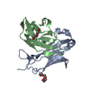
Mass: 150.173 Da / Num. of mol.: 2 / Source method: obtained synthetically / Formula: C6H14O4
#3: Water ChemComp-HOH / water


Mass: 18.015 Da / Num. of mol.: 159 / Source method: isolated from a natural source / Formula: H2O

-
Experimental details

-
Experiment

ExperimentMethod: X-RAY DIFFRACTION / Number of used crystals: 1

-
Sample preparation

CrystalDensity Matthews: 1.73 Å3/Da / Density % sol: 28.74 %
Crystal growTemperature: 295 K / Method: vapor diffusion, sitting drop / pH: 8.3
Details: Protein: 11.0 mg/ml, 0.25M Sodium chloride, 0.1M Tris HCl (pH 8.3); Screen: PEGs II (F4), 0.2M Ammonium sulfate, 0.1M Sodium acetate, 22% (w/v) PEG 4000

-
Data collection

DiffractionMean temperature: 100 K
Diffraction sourceSource: SYNCHROTRON / Site: APS / Beamline: 21-ID-D / Wavelength: 1.07809 Å
DetectorType: MARMOSAIC 300 mm CCD / Detector: CCD / Date: Aug 19, 2010 / Details: Si(111)
RadiationMonochromator: mirrors / Protocol: SINGLE WAVELENGTH / Monochromatic (M) / Laue (L): M / Scattering type: x-ray
Radiation wavelengthWavelength: 1.07809 Å / Relative weight: 1
ReflectionResolution: 1.52→30 Å / Num. obs: 31266 / % possible obs: 99.4 % / Observed criterion σ(I): -3 / Redundancy: 3.3 % / Biso Wilson estimate: 22.3 Å2 / CC1/2: 0.719 / Rmerge(I) obs: 0.055 / Rsym value: 0.055 / Net I/σ(I): 19.3
Reflection shellResolution: 1.52→1.55 Å / Redundancy: 2.9 % / Rmerge(I) obs: 0.626 / Mean I/σ(I) obs: 2.3 / % possible all: 98.5

-
Processing

Software
NameVersionClassification
REFMAC5.8.0135refinement
HKL-3000data reduction
HKL-3000data scaling
PHASERphasing
RefinementMethod to determine structure: MOLECULAR REPLACEMENT
Starting model: PDB-1HZH
Resolution: 1.52→27.44 Å / Cor.coef. Fo:Fc: 0.974 / Cor.coef. Fo:Fc free: 0.967 / SU B: 3.252 / SU ML: 0.056 / Cross valid method: THROUGHOUT / ESU R: 0.077 / ESU R Free: 0.078 / Details: HYDROGENS HAVE BEEN ADDED IN THE RIDING POSITIONS
RfactorNum. reflection% reflectionSelection details
Rfree0.18832 1570 5 %RANDOM
Rwork0.15721 ---
obs0.15875 29684 98.61 %-
Solvent computationIon probe radii: 0.6 Å / Shrinkage radii: 0.6 Å / VDW probe radii: 1.2 Å
Displacement parametersBiso mean: 28.301 Å2
Baniso -1Baniso -2Baniso -3
1-1.83 Å20 Å2-1.17 Å2
2---1.16 Å20 Å2
3----0.56 Å2
Refinement stepCycle: 1 / Resolution: 1.52→27.44 Å
ProteinNucleic acidLigandSolventTotal
Num. atoms1699 0 17 160 1876
Refine LS restraints
Refine-IDTypeDev idealDev ideal targetNumber
X-RAY DIFFRACTIONr_bond_refined_d0.010.0192026
X-RAY DIFFRACTIONr_bond_other_d0.0010.021837
X-RAY DIFFRACTIONr_angle_refined_deg1.5351.9592781
X-RAY DIFFRACTIONr_angle_other_deg0.85534287
X-RAY DIFFRACTIONr_dihedral_angle_1_deg3.2655262
X-RAY DIFFRACTIONr_dihedral_angle_2_deg31.1525.30698
X-RAY DIFFRACTIONr_dihedral_angle_3_deg9.05615340
X-RAY DIFFRACTIONr_dihedral_angle_4_deg14.063159
X-RAY DIFFRACTIONr_chiral_restr0.0990.2286
X-RAY DIFFRACTIONr_gen_planes_refined0.0210.0212414
X-RAY DIFFRACTIONr_gen_planes_other0.0170.02471
X-RAY DIFFRACTIONr_nbd_refined
X-RAY DIFFRACTIONr_nbd_other
X-RAY DIFFRACTIONr_nbtor_refined
X-RAY DIFFRACTIONr_nbtor_other
X-RAY DIFFRACTIONr_xyhbond_nbd_refined
X-RAY DIFFRACTIONr_xyhbond_nbd_other
X-RAY DIFFRACTIONr_metal_ion_refined
X-RAY DIFFRACTIONr_metal_ion_other
X-RAY DIFFRACTIONr_symmetry_vdw_refined
X-RAY DIFFRACTIONr_symmetry_vdw_other
X-RAY DIFFRACTIONr_symmetry_hbond_refined
X-RAY DIFFRACTIONr_symmetry_hbond_other
X-RAY DIFFRACTIONr_symmetry_metal_ion_refined
X-RAY DIFFRACTIONr_symmetry_metal_ion_other
X-RAY DIFFRACTIONr_mcbond_it1.5511.756988
X-RAY DIFFRACTIONr_mcbond_other1.5451.753987
X-RAY DIFFRACTIONr_mcangle_it2.5652.6181270
X-RAY DIFFRACTIONr_mcangle_other2.5642.6211271
X-RAY DIFFRACTIONr_scbond_it2.2622.0821038
X-RAY DIFFRACTIONr_scbond_other2.2612.0821038
X-RAY DIFFRACTIONr_scangle_it
X-RAY DIFFRACTIONr_scangle_other3.4972.9911512
X-RAY DIFFRACTIONr_long_range_B_refined6.25514.7482182
X-RAY DIFFRACTIONr_long_range_B_other6.1314.1262119
X-RAY DIFFRACTIONr_rigid_bond_restr
X-RAY DIFFRACTIONr_sphericity_free
X-RAY DIFFRACTIONr_sphericity_bonded
LS refinement shellResolution: 1.521→1.56 Å / Total num. of bins used: 20
RfactorNum. reflection% reflection
Rfree0.291 102 -
Rwork0.254 1973 -
obs--89.06 %
Refinement TLS params.

Method: refined / Refine-ID: X-RAY DIFFRACTION

IDL112)L122)L132)L222)L232)L332)S11 (Å °)S12 (Å °)S13 (Å °)S21 (Å °)S22 (Å °)S23 (Å °)S31 (Å °)S32 (Å °)S33 (Å °)T112)T122)T132)T222)T232)T332)Origin x (Å)Origin y (Å)Origin z (Å)
11.3297-0.6275-0.20795.00120.08822.5018-0.205-0.2440.11510.28710.1726-0.58150.1420.25740.03240.10420.0145-0.00180.13860.0020.1398-11.15553.447-7.1316
23.1811-1.1268-0.7833.39540.58331.0671-0.0080.00250.1137-0.1209-0.0074-0.09210.02930.02580.01550.03720.0070.01370.03240.00530.0109-16.16554.9827-11.6994
33.8860.78210.16874.0438-2.02787.3322-0.0003-0.073-0.0399-0.04160.09750.22470.4207-0.3688-0.09710.027-0.02450.00390.0323-0.00960.034-28.2392.262-10.0644
41.5025-0.3017-0.47611.68940.18971.0126-0.0476-0.0080.0149-0.0327-0.0058-0.08580.03350.04780.05340.01690.00810.01160.0418-0.00290.0171-17.54753.8073-11.1003
53.5749-5.4929-1.741210.93632.59751.0383-0.1521-0.19090.02590.36610.0612-0.06140.14820.06260.0910.077-0.00260.02940.0577-0.01730.0645-19.0309-1.6075-2.0093
61.387-0.51171.67390.3418-1.15424.5519-0.0795-0.05510.04250.03190.1072-0.0313-0.2164-0.2988-0.02760.0903-0.0047-0.01190.0848-0.00060.0694-18.347718.4296-18.9265
71.4416-0.69080.95621.9327-1.74073.1546-0.0763-0.0014-0.04880.04760.0086-0.178-0.11960.12640.06770.021-0.001-0.00890.03150.00120.0474-8.842511.7345-12.1826
88.3749-1.16181.81744.38330.29092.56310.00450.08970.0528-0.38970.0004-0.3872-0.26270.2896-0.0050.0595-0.03410.02380.08630.02690.06923.946518.1528-12.6987
91.18210.17120.41471.4797-0.57592.4135-0.0338-0.03450.01360.005-0.0602-0.234-0.11290.17470.0940.0062-0.0069-0.00050.05090.010.0402-6.803913.6802-14.3599
101.3727-1.013.15152.0081-4.258610.47590.0024-0.08180.04650.21590.07370.0288-0.4783-0.0771-0.0760.1835-0.03770.00910.1137-0.02340.1229-6.828724.0821-11.9942
Refinement TLS group
IDRefine-IDRefine TLS-IDAuth asym-IDAuth seq-ID
1X-RAY DIFFRACTION1A315 - 331
2X-RAY DIFFRACTION2A332 - 354
3X-RAY DIFFRACTION3A355 - 365
4X-RAY DIFFRACTION4A366 - 406
5X-RAY DIFFRACTION5A407 - 419
6X-RAY DIFFRACTION6B312 - 328
7X-RAY DIFFRACTION7B329 - 354
8X-RAY DIFFRACTION8B355 - 363
9X-RAY DIFFRACTION9B364 - 406
10X-RAY DIFFRACTION10B407 - 419

+
About Yorodumi

-
News

-
Feb 9, 2022. New format data for meta-information of EMDB entries

New format data for meta-information of EMDB entries

  • Version 3 of the EMDB header file is now the official format.
  • The previous official version 1.9 will be removed from the archive.

Related info.:EMDB header

External links:wwPDB to switch to version 3 of the EMDB data model

-
Aug 12, 2020. Covid-19 info

Covid-19 info

URL: https://pdbjlvh1.pdbj.org/emnavi/covid19.php

New page: Covid-19 featured information page in EM Navigator.

Related info.:Covid-19 info / Mar 5, 2020. Novel coronavirus structure data

+
Mar 5, 2020. Novel coronavirus structure data

Novel coronavirus structure data

Related info.:Yorodumi Speices / Aug 12, 2020. Covid-19 info

External links:COVID-19 featured content - PDBj / Molecule of the Month (242):Coronavirus Proteases

+
Jan 31, 2019. EMDB accession codes are about to change! (news from PDBe EMDB page)

EMDB accession codes are about to change! (news from PDBe EMDB page)

  • The allocation of 4 digits for EMDB accession codes will soon come to an end. Whilst these codes will remain in use, new EMDB accession codes will include an additional digit and will expand incrementally as the available range of codes is exhausted. The current 4-digit format prefixed with “EMD-” (i.e. EMD-XXXX) will advance to a 5-digit format (i.e. EMD-XXXXX), and so on. It is currently estimated that the 4-digit codes will be depleted around Spring 2019, at which point the 5-digit format will come into force.
  • The EM Navigator/Yorodumi systems omit the EMD- prefix.

Related info.:Q: What is EMD? / ID/Accession-code notation in Yorodumi/EM Navigator

External links:EMDB Accession Codes are Changing Soon! / Contact to PDBj

+
Jul 12, 2017. Major update of PDB

Major update of PDB

  • wwPDB released updated PDB data conforming to the new PDBx/mmCIF dictionary.
  • This is a major update changing the version number from 4 to 5, and with Remediation, in which all the entries are updated.
  • In this update, many items about electron microscopy experimental information are reorganized (e.g. em_software).
  • Now, EM Navigator and Yorodumi are based on the updated data.

External links:wwPDB Remediation / Enriched Model Files Conforming to OneDep Data Standards Now Available in the PDB FTP Archive

-
Yorodumi

Thousand views of thousand structures

  • Yorodumi is a browser for structure data from EMDB, PDB, SASBDB, etc.
  • This page is also the successor to EM Navigator detail page, and also detail information page/front-end page for Omokage search.
  • The word "yorodu" (or yorozu) is an old Japanese word meaning "ten thousand". "mi" (miru) is to see.

Related info.:EMDB / PDB / SASBDB / Comparison of 3 databanks / Yorodumi Search / Aug 31, 2016. New EM Navigator & Yorodumi / Yorodumi Papers / Jmol/JSmol / Function and homology information / Changes in new EM Navigator and Yorodumi

Read more