 Movie
Movie Controller

[English] 日本語
 Yorodumi
- PDB-5hrc: Crystal structure of an aspartate/glutamate racemase in complex w... -
+ Open data

- Basic information
| Entry | Database: PDB / ID: 5hrc | ||||||||||||
|---|---|---|---|---|---|---|---|---|---|---|---|---|---|
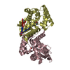
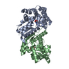
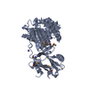
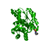
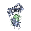
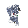
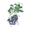
| Title | Crystal structure of an aspartate/glutamate racemase in complex with L-aspartate | ||||||||||||
 Components Components | aspartate/glutamate racemase | ||||||||||||
 Keywords Keywords | ISOMERASE / Aspartate/glutamate racemase / PLP-independent racemase / Racemization mechanism | ||||||||||||
| Function / homology | Rossmann fold - #1860 / Rossmann fold / 3-Layer(aba) Sandwich / Alpha Beta / ASPARTIC ACID / :  Function and homology information Function and homology information | ||||||||||||
| Biological species |  | ||||||||||||
| Method |  X-RAY DIFFRACTION / SYNCHROTRON / MOLECULAR REPLACEMENT / Resolution: 1.765 Å X-RAY DIFFRACTION / SYNCHROTRON / MOLECULAR REPLACEMENT / Resolution: 1.765 Å | ||||||||||||
 Authors Authors | Liu, X. / Gao, F. / Ma, Y. / Liu, S. / Cui, Y. / Yuan, Z. / Kang, X. | ||||||||||||
| Funding support |  China, 3items China, 3items
| ||||||||||||
 Citation Citation | Journal: Febs Lett. / Year: 2016 Title: Crystal structure and molecular mechanism of an aspartate/glutamate racemase from Escherichia coli O157 Authors: Liu, X. / Gao, F. / Ma, Y. / Liu, S. / Cui, Y. / Yuan, Z. / Kang, X. | ||||||||||||
| History |
|
- Structure visualization
| Structure viewer | Molecule: MolmilJmol/JSmol |
|---|
- Downloads & links
-Download
| PDBx/mmCIF format | 5hrc.cif.gz | 115.3 KB | Display | PDBx/mmCIF format |
|---|---|---|---|---|
| PDB format | pdb5hrc.ent.gz | 86.2 KB | Display | PDB format |
| PDBx/mmJSON format | 5hrc.json.gz | Tree view | PDBx/mmJSON format | |
| Others |  Other downloads Other downloads |
-Validation report
| Arichive directory | https://data.pdbj.org/pub/pdb/validation_reports/hr/5hrcftp://data.pdbj.org/pub/pdb/validation_reports/hr/5hrc | HTTPS FTP |
|---|
-Related structure data
| Related structure data |  5hqtSC  5hraC S: Starting model for refinement C: citing same article ( |
|---|---|
| Similar structure data |


-Links
 PDBj
PDBj
- Assembly
| Deposited unit | 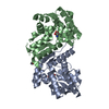
| ||||||||
|---|---|---|---|---|---|---|---|---|---|
| 1 |
| ||||||||
| Unit cell |
|
-Components
| #1: Protein | Mass: 25990.686 Da / Num. of mol.: 2 Source method: isolated from a genetically manipulated source Source: (gene. exp.) Strain: SS52 / Gene: ygeA, SS52_3985 / Plasmid: pET-21b / Production host: #2: Chemical |   Type: L-peptide linking / Mass: 133.103 Da / Num. of mol.: 2 / Source method: isolated from a natural source / Formula: C4H7NO4 Type: L-peptide linking / Mass: 133.103 Da / Num. of mol.: 2 / Source method: isolated from a natural source / Formula: C4H7NO4#3: Chemical | ChemComp-NHE / |   Mass: 207.290 Da / Num. of mol.: 1 / Source method: obtained synthetically / Formula: C8H17NO3S / Comment: pH buffer*YM Mass: 207.290 Da / Num. of mol.: 1 / Source method: obtained synthetically / Formula: C8H17NO3S / Comment: pH buffer*YM#4: Water | ChemComp-HOH / |  Mass: 18.015 Da / Num. of mol.: 504 / Source method: isolated from a natural source / Formula: H2O Mass: 18.015 Da / Num. of mol.: 504 / Source method: isolated from a natural source / Formula: H2O |
|---|
-Experimental details
-Experiment
| Experiment | Method: X-RAY DIFFRACTION / Number of used crystals: 1 |
|---|
- Sample preparation
| Crystal | Density Matthews: 2.51 Å3/Da / Density % sol: 50.95 % |
|---|---|
| Crystal grow | Temperature: 293 K / Method: vapor diffusion / pH: 9.5 Details: 1.0 M sodium citrate, 0.1 M CHES, 3.0% D (+)-Sucrose |
-Data collection
| Diffraction | Mean temperature: 100 K |
|---|---|
| Diffraction source | Source: SYNCHROTRON / Site: SSRF / Beamline: BL17U / Wavelength: 0.989 Å |
| Detector | Type: ADSC QUANTUM 315r / Detector: CCD / Date: Oct 12, 2015 |
| Radiation | Protocol: SINGLE WAVELENGTH / Monochromatic (M) / Laue (L): M / Scattering type: x-ray |
| Radiation wavelength | Wavelength: 0.989 Å / Relative weight: 1 |
| Reflection | Resolution: 1.765→32.73 Å / Num. obs: 48432 / % possible obs: 91 % / Redundancy: 3.4 % / Net I/σ(I): 20.06 |
| Reflection shell | Resolution: 1.765→1.828 Å |
- Processing
| Software |
| ||||||||||||||||||||||||||||||||||||||||||||||||||||||||||||||||||||||||||||||||||||||||||||||||||||||||||||||||||||||||||||||
|---|---|---|---|---|---|---|---|---|---|---|---|---|---|---|---|---|---|---|---|---|---|---|---|---|---|---|---|---|---|---|---|---|---|---|---|---|---|---|---|---|---|---|---|---|---|---|---|---|---|---|---|---|---|---|---|---|---|---|---|---|---|---|---|---|---|---|---|---|---|---|---|---|---|---|---|---|---|---|---|---|---|---|---|---|---|---|---|---|---|---|---|---|---|---|---|---|---|---|---|---|---|---|---|---|---|---|---|---|---|---|---|---|---|---|---|---|---|---|---|---|---|---|---|---|---|---|---|
| Refinement | Method to determine structure: MOLECULAR REPLACEMENT Starting model: 5HQT Resolution: 1.765→32.73 Å / SU ML: 0.22 / Cross valid method: FREE R-VALUE / σ(F): 0 / Phase error: 27.26 / Stereochemistry target values: ML
| ||||||||||||||||||||||||||||||||||||||||||||||||||||||||||||||||||||||||||||||||||||||||||||||||||||||||||||||||||||||||||||||
| Solvent computation | Shrinkage radii: 0.9 Å / VDW probe radii: 1.11 Å / Solvent model: FLAT BULK SOLVENT MODEL | ||||||||||||||||||||||||||||||||||||||||||||||||||||||||||||||||||||||||||||||||||||||||||||||||||||||||||||||||||||||||||||||
| Refinement step | Cycle: LAST / Resolution: 1.765→32.73 Å
| ||||||||||||||||||||||||||||||||||||||||||||||||||||||||||||||||||||||||||||||||||||||||||||||||||||||||||||||||||||||||||||||
| Refine LS restraints |
| ||||||||||||||||||||||||||||||||||||||||||||||||||||||||||||||||||||||||||||||||||||||||||||||||||||||||||||||||||||||||||||||
| LS refinement shell |
|
