 Movie
Movie Controller


+ Open data

- Basic information
| Entry | Database: PDB / ID: 5hns | |||||||||
|---|---|---|---|---|---|---|---|---|---|---|
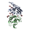
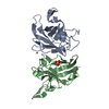
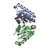
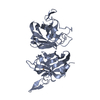
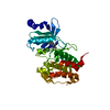
| Title | Structure of glycosylated NPC1 luminal domain C | |||||||||
 Components Components | Niemann-Pick C1 protein | |||||||||
 Keywords Keywords | PROTEIN BINDING / Niemann-Pick disease type C / NPC1 / NPC2 / cholesterol transport / Ebola virus receptor / Ebola virus susceptibility | |||||||||
| Function / homology |  Function and homology information Function and homology informationmembrane raft organization / cytoplasmic side of lysosomal membrane / intracellular cholesterol transport / intracellular lipid transport / lysosome to ER cholesterol transport / negative regulation of epithelial cell apoptotic process / sterol transport / intestinal cholesterol absorption / cholesterol transport / bile acid metabolic process ...membrane raft organization / cytoplasmic side of lysosomal membrane / intracellular cholesterol transport / intracellular lipid transport / lysosome to ER cholesterol transport / negative regulation of epithelial cell apoptotic process / sterol transport / intestinal cholesterol absorption / cholesterol transport / bile acid metabolic process / LDL clearance / cholesterol transfer activity / establishment of protein localization to membrane / glycoprotein biosynthetic process / cholesterol efflux / lysosomal transport / cholesterol binding / response to cadmium ion / cholesterol metabolic process / negative regulation of TORC1 signaling / cholesterol homeostasis / gene expression / autophagy / nuclear envelope / transmembrane signaling receptor activity / late endosome membrane / virus receptor activity / signaling receptor activity / lysosome / membrane raft / lysosomal membrane / symbiont entry into host cell / perinuclear region of cytoplasm / Golgi apparatus / endoplasmic reticulum / extracellular exosome / extracellular region / membrane / plasma membrane Similarity search - Function | |||||||||
| Biological species |  Homo sapiens (human) Homo sapiens (human) | |||||||||
| Method |  X-RAY DIFFRACTION / SYNCHROTRON / SAD / Resolution: 2.45 Å X-RAY DIFFRACTION / SYNCHROTRON / SAD / Resolution: 2.45 Å | |||||||||
 Authors Authors | Zhao, Y. / Ren, J. / Harlos, K. / Stuart, D.I. | |||||||||
 Citation Citation | Journal: Febs Lett. / Year: 2016 Title: Structure of glycosylated NPC1 luminal domain C reveals insights into NPC2 and Ebola virus interactions. Authors: Zhao, Y. / Ren, J. / Harlos, K. / Stuart, D.I. | |||||||||
| History |
|
- Structure visualization
| Structure viewer | Molecule: MolmilJmol/JSmol |
|---|
- Downloads & links
-Download
| PDBx/mmCIF format | 5hns.cif.gz | 201.2 KB | Display | PDBx/mmCIF format |
|---|---|---|---|---|
| PDB format | pdb5hns.ent.gz | 164 KB | Display | PDB format |
| PDBx/mmJSON format | 5hns.json.gz | Tree view | PDBx/mmJSON format | |
| Others |  Other downloads Other downloads |
-Validation report
| Arichive directory | https://data.pdbj.org/pub/pdb/validation_reports/hn/5hnsftp://data.pdbj.org/pub/pdb/validation_reports/hn/5hns | HTTPS FTP |
|---|
-Related structure data
| Similar structure data |
|---|



-Links
 PDBj
PDBj
- Assembly
| Deposited unit | 
| ||||||||||||||||||
|---|---|---|---|---|---|---|---|---|---|---|---|---|---|---|---|---|---|---|---|
| 1 | 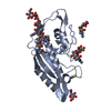
| ||||||||||||||||||
| 2 | 
| ||||||||||||||||||
| Unit cell |
| ||||||||||||||||||
| Noncrystallographic symmetry (NCS) | NCS domain:
NCS domain segments: Component-ID: _ / Ens-ID: 1 / Beg auth comp-ID: LYS / Beg label comp-ID: LYS / End auth comp-ID: ALA / End label comp-ID: ALA / Refine code: _ / Auth seq-ID: 392 - 605 / Label seq-ID: 9 - 222
|
-Components
-Protein , 1 types, 2 molecules AB
| #1: Protein | Mass: 27952.037 Da / Num. of mol.: 2 / Fragment: domain C, UNP residues 387-618 Source method: isolated from a genetically manipulated source Source: (gene. exp.) Homo sapiens (human) / Gene: NPC1 / Cell line (production host): HEK293S / Organ (production host): Kidney / Production host: Homo sapiens (human) / References: UniProt: O15118 |
|---|
-Sugars , 5 types, 9 molecules 
| #2: Polysaccharide | 2-acetamido-2-deoxy-beta-D-glucopyranose-(1-4)-2-acetamido-2-deoxy-beta-D-glucopyranose Source method: isolated from a genetically manipulated source Source: (gene. exp.) Homo sapiens (human) / Cell line (production host): HEK293S / Organ (production host): kidney / Production host: Homo sapiens (human)#3: Polysaccharide | alpha-D-mannopyranose-(1-6)-beta-D-mannopyranose-(1-4)-2-acetamido-2-deoxy-beta-D-glucopyranose-(1- ...alpha-D-mannopyranose-(1-6)-beta-D-mannopyranose-(1-4)-2-acetamido-2-deoxy-beta-D-glucopyranose-(1-4)-2-acetamido-2-deoxy-beta-D-glucopyranose | Source method: isolated from a genetically manipulated source Source: (gene. exp.) Homo sapiens (human) / Cell line (production host): HEK293S / Organ (production host): kidney / Production host: Homo sapiens (human)#4: Polysaccharide | alpha-D-mannopyranose-(1-3)-[alpha-D-mannopyranose-(1-6)]beta-D-mannopyranose-(1-4)-2-acetamido-2- ...alpha-D-mannopyranose-(1-3)-[alpha-D-mannopyranose-(1-6)]beta-D-mannopyranose-(1-4)-2-acetamido-2-deoxy-beta-D-glucopyranose-(1-4)-2-acetamido-2-deoxy-beta-D-glucopyranose | Source method: isolated from a genetically manipulated source #5: Polysaccharide | beta-D-mannopyranose-(1-4)-2-acetamido-2-deoxy-beta-D-glucopyranose-(1-4)-2-acetamido-2-deoxy-beta- ...beta-D-mannopyranose-(1-4)-2-acetamido-2-deoxy-beta-D-glucopyranose-(1-4)-2-acetamido-2-deoxy-beta-D-glucopyranose | Source method: isolated from a genetically manipulated source Source: (gene. exp.) Homo sapiens (human) / Cell line (production host): HEK293S / Organ (production host): kidney / Production host: Homo sapiens (human)#6: Sugar | ChemComp-NAG / |  Type: D-saccharide, beta linking / Mass: 221.208 Da / Num. of mol.: 1 Type: D-saccharide, beta linking / Mass: 221.208 Da / Num. of mol.: 1Source method: isolated from a genetically manipulated source Formula: C8H15NO6 / Source: (gene. exp.) Homo sapiens (human) / Cell line (production host): HEK293S / Organ (production host): kidney / Production host: Homo sapiens (human) |
|---|
-Non-polymers , 2 types, 48 molecules 

| #7: Chemical | ChemComp-SCN / 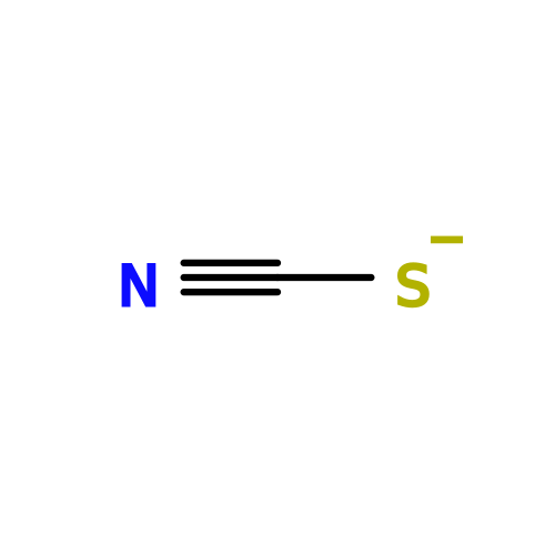 Mass: 58.082 Da / Num. of mol.: 4 Mass: 58.082 Da / Num. of mol.: 4Source method: isolated from a genetically manipulated source Formula: CNS #8: Water | ChemComp-HOH / | Mass: 18.015 Da / Num. of mol.: 44 / Source method: isolated from a natural source / Formula: H2O |
|---|
-Details
| Has protein modification | Y |
|---|
-Experimental details
-Experiment
| Experiment | Method: X-RAY DIFFRACTION / Number of used crystals: 1 |
|---|
- Sample preparation
| Crystal | Density Matthews: 3.36 Å3/Da / Density % sol: 63.37 % |
|---|---|
| Crystal grow | Temperature: 293 K / Method: vapor diffusion, sitting drop / pH: 7.4 Details: 30% polyethylene glycol mono-ethyl Ether 2000 and 0.1 M potassium thiocyanate |
-Data collection
| Diffraction | Mean temperature: 100 K |
|---|---|
| Diffraction source | Source: SYNCHROTRON / Site: Diamond  / Beamline: I04 / Wavelength: 1.0675 Å / Beamline: I04 / Wavelength: 1.0675 Å |
| Detector | Type: DECTRIS PILATUS 6M / Detector: PIXEL / Date: Jul 18, 2015 |
| Radiation | Protocol: SINGLE WAVELENGTH / Monochromatic (M) / Laue (L): M / Scattering type: x-ray |
| Radiation wavelength | Wavelength: 1.0675 Å / Relative weight: 1 |
| Reflection | Resolution: 2.45→70 Å / Num. obs: 28062 / % possible obs: 100 % / Redundancy: 26.5 % / Net I/σ(I): 13.3 |
- Processing
| Software |
| ||||||||||||||||||||||||||||||||||||||||||||||||||||||||||||||||||||||||||||||||||||||||||||||||||||||||||||||||||||||||||||||||||||||||||||||||||||||||||||||||||||||||||||||||||||||
|---|---|---|---|---|---|---|---|---|---|---|---|---|---|---|---|---|---|---|---|---|---|---|---|---|---|---|---|---|---|---|---|---|---|---|---|---|---|---|---|---|---|---|---|---|---|---|---|---|---|---|---|---|---|---|---|---|---|---|---|---|---|---|---|---|---|---|---|---|---|---|---|---|---|---|---|---|---|---|---|---|---|---|---|---|---|---|---|---|---|---|---|---|---|---|---|---|---|---|---|---|---|---|---|---|---|---|---|---|---|---|---|---|---|---|---|---|---|---|---|---|---|---|---|---|---|---|---|---|---|---|---|---|---|---|---|---|---|---|---|---|---|---|---|---|---|---|---|---|---|---|---|---|---|---|---|---|---|---|---|---|---|---|---|---|---|---|---|---|---|---|---|---|---|---|---|---|---|---|---|---|---|---|---|
| Refinement | Method to determine structure: SAD / Resolution: 2.45→70.03 Å / Cor.coef. Fo:Fc: 0.946 / Cor.coef. Fo:Fc free: 0.941 / SU B: 21.463 / SU ML: 0.219 / Cross valid method: THROUGHOUT / ESU R: 0.336 / ESU R Free: 0.228 / Stereochemistry target values: MAXIMUM LIKELIHOOD / Details: HYDROGENS HAVE BEEN ADDED IN THE RIDING POSITIONS
| ||||||||||||||||||||||||||||||||||||||||||||||||||||||||||||||||||||||||||||||||||||||||||||||||||||||||||||||||||||||||||||||||||||||||||||||||||||||||||||||||||||||||||||||||||||||
| Solvent computation | Ion probe radii: 0.8 Å / Shrinkage radii: 0.8 Å / VDW probe radii: 1.2 Å / Solvent model: MASK | ||||||||||||||||||||||||||||||||||||||||||||||||||||||||||||||||||||||||||||||||||||||||||||||||||||||||||||||||||||||||||||||||||||||||||||||||||||||||||||||||||||||||||||||||||||||
| Displacement parameters | Biso mean: 73.014 Å2
| ||||||||||||||||||||||||||||||||||||||||||||||||||||||||||||||||||||||||||||||||||||||||||||||||||||||||||||||||||||||||||||||||||||||||||||||||||||||||||||||||||||||||||||||||||||||
| Refinement step | Cycle: LAST / Resolution: 2.45→70.03 Å
| ||||||||||||||||||||||||||||||||||||||||||||||||||||||||||||||||||||||||||||||||||||||||||||||||||||||||||||||||||||||||||||||||||||||||||||||||||||||||||||||||||||||||||||||||||||||
| Refine LS restraints |
|