- PDB-3qtb: Structure of the universal stress protein from Archaeoglobus fulg... -
+
Open data
ID or keywords:
Loading...
-
Basic information
Entry
Database: PDB / ID: 3qtb
Title
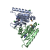
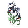
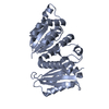
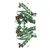
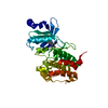
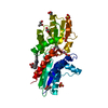
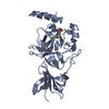
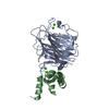
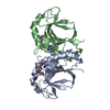
Structure of the universal stress protein from Archaeoglobus fulgidus in complex with dAMP
Components
Uncharacterized protein
Keywords
STRUCTURAL GENOMICS / UNKNOWN FUNCTION / PSI-Biology / Midwest Center for Structural Genomics / MCSG / a/b class / RFM like / putative universal stress protein
Function / homology
Function and homology information
Universal stress protein A family / UspA / Universal stress protein family / HUPs / Rossmann-like alpha/beta/alpha sandwich fold / Rossmann fold / 3-Layer(aba) Sandwich / Alpha Beta Similarity search - Domain/homology
Resolution: 2.1→50 Å / Cor.coef. Fo:Fc: 0.951 / Cor.coef. Fo:Fc free: 0.936 / SU B: 12.228 / SU ML: 0.153 / Cross valid method: THROUGHOUT / σ(F): 0 / σ(I): 0 / ESU R: 0.254 / ESU R Free: 0.195 / Stereochemistry target values: MAXIMUM LIKELIHOOD / Details: HYDROGENS HAVE BEEN USED IF PRESENT IN THE INPUT
Rfactor
Num. reflection
% reflection
Selection details
Rfree
0.23747
738
5 %
RANDOM
Rwork
0.1997
-
-
-
all
0.2017
14059
-
-
obs
0.2017
14059
98.78 %
-
Solvent computation
Ion probe radii: 0.8 Å / Shrinkage radii: 0.8 Å / VDW probe radii: 1.2 Å / Solvent model: BABINET MODEL WITH MASK
Displacement parameters
Biso mean: 38.86 Å2
Baniso -1
Baniso -2
Baniso -3
1-
-0.1 Å2
0 Å2
-1.35 Å2
2-
-
0.63 Å2
0 Å2
3-
-
-
-1.75 Å2
Refinement step
Cycle: LAST / Resolution: 2.1→50 Å
Protein
Nucleic acid
Ligand
Solvent
Total
Num. atoms
1970
0
34
50
2054
Refine LS restraints
Refine-ID
Type
Dev ideal
Dev ideal target
Number
X-RAY DIFFRACTION
r_bond_refined_d
0.014
0.022
2031
X-RAY DIFFRACTION
r_bond_other_d
0.006
0.02
1394
X-RAY DIFFRACTION
r_angle_refined_deg
1.425
2.003
2750
X-RAY DIFFRACTION
r_angle_other_deg
0.84
3
3402
X-RAY DIFFRACTION
r_dihedral_angle_1_deg
5.673
5
255
X-RAY DIFFRACTION
r_dihedral_angle_2_deg
34.469
23.333
81
X-RAY DIFFRACTION
r_dihedral_angle_3_deg
15.158
15
355
X-RAY DIFFRACTION
r_dihedral_angle_4_deg
15.662
15
19
X-RAY DIFFRACTION
r_chiral_restr
0.081
0.2
323
X-RAY DIFFRACTION
r_gen_planes_refined
0.005
0.021
2228
X-RAY DIFFRACTION
r_gen_planes_other
0.001
0.02
388
LS refinement shell
Resolution: 2.1→2.154 Å / Total num. of bins used: 20
Rfactor
Num. reflection
% reflection
Rfree
0.218
54
-
Rwork
0.196
1027
-
obs
-
-
99.45 %
Refinement TLS params.
Method: refined / Refine-ID: X-RAY DIFFRACTION
ID
L11 (°2)
L12 (°2)
L13 (°2)
L22 (°2)
L23 (°2)
L33 (°2)
S11 (Å °)
S12 (Å °)
S13 (Å °)
S21 (Å °)
S22 (Å °)
S23 (Å °)
S31 (Å °)
S32 (Å °)
S33 (Å °)
T11 (Å2)
T12 (Å2)
T13 (Å2)
T22 (Å2)
T23 (Å2)
T33 (Å2)
Origin x (Å)
Origin y (Å)
Origin z (Å)
1
2.8696
0.7256
-0.8763
1.2427
-0.3633
1.3092
-0.0394
-0.0858
-0.0144
0.0149
0.0671
0.0196
-0.0344
0.1352
-0.0276
0.074
0.0267
-0.0219
0.0374
-0.0078
0.0524
19.7546
29.7929
-13.3028
2
6.8741
-6.571
-1.898
10.1758
6.4603
13.7842
0.315
0.4575
-0.0253
-0.3762
-0.1049
-0.2151
-0.0372
0.196
-0.2102
0.0766
0.0188
-0.0007
0.1751
-0.0343
0.1278
29.2668
23.4723
-29.6174
3
3.2047
0.9418
-1.6701
1.7717
-0.1296
1.0569
-0.09
-0.1662
-0.253
0.0793
0.0851
-0.085
0.1208
0.1892
0.0048
0.1685
0.0141
-0.031
0.1739
-0.0282
0.196
24.1643
23.0176
-15.2641
Refinement TLS group
ID
Refine-ID
Refine TLS-ID
Auth asym-ID
Auth seq-ID
1
X-RAY DIFFRACTION
1
A
1 - 104
2
X-RAY DIFFRACTION
1
B
1 - 102
3
X-RAY DIFFRACTION
2
B
103 - 116
4
X-RAY DIFFRACTION
3
A
116 - 134
5
X-RAY DIFFRACTION
3
B
117 - 134
+
About Yorodumi
-
News
-
Feb 9, 2022. New format data for meta-information of EMDB entries
New format data for meta-information of EMDB entries
Version 3 of the EMDB header file is now the official format.
The previous official version 1.9 will be removed from the archive.
In the structure databanks used in Yorodumi, some data are registered as the other names, "COVID-19 virus" and "2019-nCoV". Here are the details of the virus and the list of structure data.
Jan 31, 2019. EMDB accession codes are about to change! (news from PDBe EMDB page)
EMDB accession codes are about to change! (news from PDBe EMDB page)
The allocation of 4 digits for EMDB accession codes will soon come to an end. Whilst these codes will remain in use, new EMDB accession codes will include an additional digit and will expand incrementally as the available range of codes is exhausted. The current 4-digit format prefixed with “EMD-” (i.e. EMD-XXXX) will advance to a 5-digit format (i.e. EMD-XXXXX), and so on. It is currently estimated that the 4-digit codes will be depleted around Spring 2019, at which point the 5-digit format will come into force.
The EM Navigator/Yorodumi systems omit the EMD- prefix.
Related info.:Q: What is EMD? / ID/Accession-code notation in Yorodumi/EM Navigator
Yorodumi is a browser for structure data from EMDB, PDB, SASBDB, etc.
This page is also the successor to EM Navigator detail page, and also detail information page/front-end page for Omokage search.
The word "yorodu" (or yorozu) is an old Japanese word meaning "ten thousand". "mi" (miru) is to see.
Related info.:EMDB / PDB / SASBDB / Comparison of 3 databanks / Yorodumi Search / Aug 31, 2016. New EM Navigator & Yorodumi / Yorodumi Papers / Jmol/JSmol / Function and homology information / Changes in new EM Navigator and Yorodumi
 Movie
Movie Controller

 Yorodumi
 Open data
 Basic information
 Components
 Keywords
 Function and homology information
 Archaeoglobus fulgidus (archaea)
 X-RAY DIFFRACTION /
  Authors
 Citation
 Structure visualization
 Downloads & links
 Other downloads
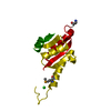


 PDBj
PDBj Assembly
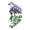

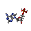
 Mass: 331.222 Da / Num. of mol.: 1 / Source method: obtained synthetically / Formula: C10H14N5O6P
 Mass: 59.044 Da / Num. of mol.: 3 / Source method: obtained synthetically / Formula: C2H3O2
 Mass: 18.015 Da / Num. of mol.: 50 / Source method: isolated from a natural source / Formula: H2O
 Sample preparation
 / Beamline: 21-ID-G / Wavelength: 0.9786 Å
 Processing