 Movie
Movie Controller

[English] 日本語
 Yorodumi
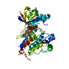
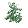
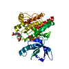
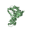
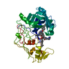
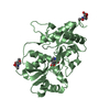
- PDB-5hml: Crystal Structure of T5 D15 Protein Co-crystallized with Metal Ions -
+ Open data

- Basic information
| Entry | Database: PDB / ID: 5hml | ||||||
|---|---|---|---|---|---|---|---|
| Title | Crystal Structure of T5 D15 Protein Co-crystallized with Metal Ions | ||||||
 Components Components | Exodeoxyribonuclease | ||||||
 Keywords Keywords | HYDROLASE / Metal ion complex / flap endonuclease / alternative conformations | ||||||
| Function / homology |  Function and homology information Function and homology informationviral replication complex / exodeoxyribonuclease (lambda-induced) / late viral transcription / DNA replication, Okazaki fragment processing / double-stranded DNA 5'-3' DNA exonuclease activity / double-stranded DNA endonuclease activity / DNA exonuclease activity / 5'-flap endonuclease activity / viral DNA genome replication / 5'-3' exonuclease activity ...viral replication complex / exodeoxyribonuclease (lambda-induced) / late viral transcription / DNA replication, Okazaki fragment processing / double-stranded DNA 5'-3' DNA exonuclease activity / double-stranded DNA endonuclease activity / DNA exonuclease activity / 5'-flap endonuclease activity / viral DNA genome replication / 5'-3' exonuclease activity / 5'-3' DNA exonuclease activity / Hydrolases; Acting on ester bonds; Exodeoxyribonucleases producing 5'-phosphomonoesters / DNA binding / metal ion binding Similarity search - Function | ||||||
| Biological species |  Escherichia phage T5 (virus) Escherichia phage T5 (virus) | ||||||
| Method |  X-RAY DIFFRACTION / SYNCHROTRON / MOLECULAR REPLACEMENT / Resolution: 1.482 Å X-RAY DIFFRACTION / SYNCHROTRON / MOLECULAR REPLACEMENT / Resolution: 1.482 Å | ||||||
 Authors Authors | Flemming, C.S. / Feng, M. / Sedelnikova, S.E. / Zhang, J. / Rafferty, J.B. / Sayers, J.R. / Artymiuk, P.J. | ||||||
 Citation Citation | Journal: Nat.Struct.Mol.Biol. / Year: 2016 Title: Direct observation of DNA threading in flap endonuclease complexes. Authors: AlMalki, F.A. / Flemming, C.S. / Zhang, J. / Feng, M. / Sedelnikova, S.E. / Ceska, T. / Rafferty, J.B. / Sayers, J.R. / Artymiuk, P.J. | ||||||
| History |
|
- Structure visualization
| Structure viewer | Molecule: MolmilJmol/JSmol |
|---|
- Downloads & links
-Download
| PDBx/mmCIF format | 5hml.cif.gz | 261.2 KB | Display | PDBx/mmCIF format |
|---|---|---|---|---|
| PDB format | pdb5hml.ent.gz | 209 KB | Display | PDB format |
| PDBx/mmJSON format | 5hml.json.gz | Tree view | PDBx/mmJSON format | |
| Others |  Other downloads Other downloads |
-Validation report
| Arichive directory | https://data.pdbj.org/pub/pdb/validation_reports/hm/5hmlftp://data.pdbj.org/pub/pdb/validation_reports/hm/5hml | HTTPS FTP |
|---|
-Related structure data
| Related structure data |  5hmmC 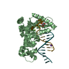 5hnkC 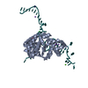 5hp4C 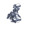 1ut5S S: Starting model for refinement C: citing same article ( |
|---|---|
| Similar structure data |


-Links
 PDBj
PDBj

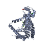
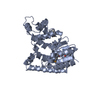
- Assembly
| Deposited unit | 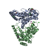
| ||||||||
|---|---|---|---|---|---|---|---|---|---|
| 1 | 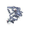
| ||||||||
| 2 | 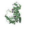
| ||||||||
| Unit cell |
|
-Components
-Protein , 1 types, 2 molecules AB
| #1: Protein | Mass: 31275.475 Da / Num. of mol.: 2 / Fragment: UNP residues 20-291 / Mutation: D134K Source method: isolated from a genetically manipulated source Details: Structure derived from recombinant protein corresponding to residues 20 - 291 (known as delta19 and had a D153K mutation compared to wt. Source: (gene. exp.) Escherichia phage T5 (virus) / Gene: D15 / Plasmid: pJONEX4Details (production host): pUC19 derivative with a lambda promoter/operator system Production host:  References: UniProt: P06229, exodeoxyribonuclease (lambda-induced) |
|---|
-Non-polymers , 6 types, 579 molecules 


| #2: Chemical |  Mass: 24.305 Da / Num. of mol.: 3 / Source method: obtained synthetically / Formula: Mg Mass: 24.305 Da / Num. of mol.: 3 / Source method: obtained synthetically / Formula: Mg#3: Chemical | ChemComp-CL /  Mass: 35.453 Da / Num. of mol.: 12 / Source method: obtained synthetically / Formula: Cl Mass: 35.453 Da / Num. of mol.: 12 / Source method: obtained synthetically / Formula: Cl#4: Chemical |  Mass: 62.068 Da / Num. of mol.: 2 / Source method: obtained synthetically / Formula: C2H6O2 Mass: 62.068 Da / Num. of mol.: 2 / Source method: obtained synthetically / Formula: C2H6O2#5: Chemical | ChemComp-B3P / |  Mass: 282.334 Da / Num. of mol.: 1 / Source method: obtained synthetically / Formula: C11H26N2O6 / Comment: pH buffer*YM Mass: 282.334 Da / Num. of mol.: 1 / Source method: obtained synthetically / Formula: C11H26N2O6 / Comment: pH buffer*YM#6: Chemical |  Mass: 126.904 Da / Num. of mol.: 2 / Source method: obtained synthetically / Formula: I Mass: 126.904 Da / Num. of mol.: 2 / Source method: obtained synthetically / Formula: I#7: Water | ChemComp-HOH / | Mass: 18.015 Da / Num. of mol.: 559 / Source method: isolated from a natural source / Formula: H2O |
|---|
-Experimental details
-Experiment
| Experiment | Method: X-RAY DIFFRACTION / Number of used crystals: 1 |
|---|
- Sample preparation
| Crystal | Density Matthews: 2.33 Å3/Da / Density % sol: 47.15 % |
|---|---|
| Crystal grow | Temperature: 280 K / Method: vapor diffusion, hanging drop / pH: 8 / Details: Bis-Tris propane, PEG 3350, MgCl2, NaI / PH range: 7.5-8.0 |
-Data collection
| Diffraction | Mean temperature: 100 K | ||||||||||||||||||||||||||||||||||||||||||||||||||||||||||||||||||||||||||||||||||||||||||||||||||||||||||||||
|---|---|---|---|---|---|---|---|---|---|---|---|---|---|---|---|---|---|---|---|---|---|---|---|---|---|---|---|---|---|---|---|---|---|---|---|---|---|---|---|---|---|---|---|---|---|---|---|---|---|---|---|---|---|---|---|---|---|---|---|---|---|---|---|---|---|---|---|---|---|---|---|---|---|---|---|---|---|---|---|---|---|---|---|---|---|---|---|---|---|---|---|---|---|---|---|---|---|---|---|---|---|---|---|---|---|---|---|---|---|---|---|
| Diffraction source | Source: SYNCHROTRON / Site: Diamond  / Beamline: I03 / Wavelength: 0.9795 Å / Beamline: I03 / Wavelength: 0.9795 Å | ||||||||||||||||||||||||||||||||||||||||||||||||||||||||||||||||||||||||||||||||||||||||||||||||||||||||||||||
| Detector | Type: DECTRIS PILATUS 6M / Detector: PIXEL / Date: Oct 10, 2010 | ||||||||||||||||||||||||||||||||||||||||||||||||||||||||||||||||||||||||||||||||||||||||||||||||||||||||||||||
| Radiation | Protocol: SINGLE WAVELENGTH / Monochromatic (M) / Laue (L): M / Scattering type: x-ray | ||||||||||||||||||||||||||||||||||||||||||||||||||||||||||||||||||||||||||||||||||||||||||||||||||||||||||||||
| Radiation wavelength | Wavelength: 0.9795 Å / Relative weight: 1 | ||||||||||||||||||||||||||||||||||||||||||||||||||||||||||||||||||||||||||||||||||||||||||||||||||||||||||||||
| Reflection | Resolution: 1.482→54.602 Å / Num. all: 87441 / Num. obs: 87441 / % possible obs: 93.7 % / Redundancy: 3 % / Rpim(I) all: 0.039 / Rrim(I) all: 0.069 / Rsym value: 0.057 / Net I/av σ(I): 10.07 / Net I/σ(I): 11.4 / Num. measured all: 264840 | ||||||||||||||||||||||||||||||||||||||||||||||||||||||||||||||||||||||||||||||||||||||||||||||||||||||||||||||
| Reflection shell | Diffraction-ID: 1 / Rejects: _
|
- Processing
| Software |
| ||||||||||||||||||||||||||||||||||||||||||||||||||||||||||||||||||||||||||||||||||||||||||
|---|---|---|---|---|---|---|---|---|---|---|---|---|---|---|---|---|---|---|---|---|---|---|---|---|---|---|---|---|---|---|---|---|---|---|---|---|---|---|---|---|---|---|---|---|---|---|---|---|---|---|---|---|---|---|---|---|---|---|---|---|---|---|---|---|---|---|---|---|---|---|---|---|---|---|---|---|---|---|---|---|---|---|---|---|---|---|---|---|---|---|---|
| Refinement | Method to determine structure: MOLECULAR REPLACEMENT Starting model: 1UT5 Resolution: 1.482→54.6 Å / Cor.coef. Fo:Fc: 0.974 / Cor.coef. Fo:Fc free: 0.954 / WRfactor Rfree: 0.1968 / WRfactor Rwork: 0.1415 / FOM work R set: 0.8709 / SU B: 3.215 / SU ML: 0.052 / SU R Cruickshank DPI: 0.0793 / SU Rfree: 0.0743 / Cross valid method: THROUGHOUT / σ(F): 0 / ESU R: 0.079 / ESU R Free: 0.074 / Stereochemistry target values: MAXIMUM LIKELIHOOD Details: HYDROGENS HAVE BEEN ADDED IN THE RIDING POSITIONS U VALUES : REFINED INDIVIDUALLY
| ||||||||||||||||||||||||||||||||||||||||||||||||||||||||||||||||||||||||||||||||||||||||||
| Solvent computation | Ion probe radii: 0.8 Å / Shrinkage radii: 0.8 Å / VDW probe radii: 1.2 Å / Solvent model: MASK | ||||||||||||||||||||||||||||||||||||||||||||||||||||||||||||||||||||||||||||||||||||||||||
| Displacement parameters | Biso max: 136.3 Å2 / Biso mean: 19.889 Å2 / Biso min: 6.82 Å2
| ||||||||||||||||||||||||||||||||||||||||||||||||||||||||||||||||||||||||||||||||||||||||||
| Refinement step | Cycle: final / Resolution: 1.482→54.6 Å
| ||||||||||||||||||||||||||||||||||||||||||||||||||||||||||||||||||||||||||||||||||||||||||
| Refine LS restraints |
| ||||||||||||||||||||||||||||||||||||||||||||||||||||||||||||||||||||||||||||||||||||||||||
| LS refinement shell | Resolution: 1.482→1.52 Å / Total num. of bins used: 20
|
