 Movie
Movie Controller

[English] 日本語
 Yorodumi
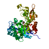
- PDB-5hmf: Crystal structure of triazine hydrolase variant (P214T/Y215H/E241Q) -
+ Open data

- Basic information
| Entry | Database: PDB / ID: 5hmf | ||||||
|---|---|---|---|---|---|---|---|
| Title | Crystal structure of triazine hydrolase variant (P214T/Y215H/E241Q) | ||||||
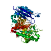
 Components Components | Triazine hydrolase | ||||||
 Keywords Keywords | HYDROLASE / amidohydrolase | ||||||
| Function / homology |  Function and homology information Function and homology informationamidase activity / hydrolase activity, acting on carbon-nitrogen (but not peptide) bonds / metal ion binding Similarity search - Function | ||||||
| Biological species |  Arthrobacter aurescens (bacteria) Arthrobacter aurescens (bacteria) | ||||||
| Method |  X-RAY DIFFRACTION / SYNCHROTRON / MOLECULAR REPLACEMENT / Resolution: 1.84 Å X-RAY DIFFRACTION / SYNCHROTRON / MOLECULAR REPLACEMENT / Resolution: 1.84 Å | ||||||
 Authors Authors | Sugrue, E. / Carr, P.D. / Jackson, C.J. | ||||||
 Citation Citation | Journal: Biochemistry / Year: 2016 Title: Active Site Desolvation and Thermostability Trade-Offs in the Evolution of Catalytically Diverse Triazine Hydrolases. Authors: Sugrue, E. / Carr, P.D. / Scott, C. / Jackson, C.J. | ||||||
| History |
|
- Structure visualization
| Structure viewer | Molecule: MolmilJmol/JSmol |
|---|
- Downloads & links
-Download
| PDBx/mmCIF format | 5hmf.cif.gz | 354.2 KB | Display | PDBx/mmCIF format |
|---|---|---|---|---|
| PDB format | pdb5hmf.ent.gz | 287.3 KB | Display | PDB format |
| PDBx/mmJSON format | 5hmf.json.gz | Tree view | PDBx/mmJSON format | |
| Others |  Other downloads Other downloads |
-Validation report
| Arichive directory | https://data.pdbj.org/pub/pdb/validation_reports/hm/5hmfftp://data.pdbj.org/pub/pdb/validation_reports/hm/5hmf | HTTPS FTP |
|---|
-Related structure data
| Related structure data |  5hmdC  5hmeC  4lh8S S: Starting model for refinement C: citing same article ( |
|---|---|
| Similar structure data |


-Links
 PDBj
PDBj

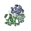
- Assembly
| Deposited unit | 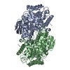
| ||||||||
|---|---|---|---|---|---|---|---|---|---|
| 1 | 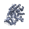
| ||||||||
| 2 | 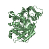
| ||||||||
| 3 |
| ||||||||
| Unit cell |
|
-Components
| #1: Protein | Mass: 50096.922 Da / Num. of mol.: 2 / Mutation: P216T, Y217H, E243Q Source method: isolated from a genetically manipulated source Source: (gene. exp.) Arthrobacter aurescens (bacteria) / Strain: TC1 / Gene: TRZN / Plasmid: petMCSIII / Production host: References: UniProt: Q6SJY7, UniProt: A1RCJ9*PLUS, atrazine chlorohydrolase #2: Chemical |   Mass: 65.409 Da / Num. of mol.: 2 / Source method: obtained synthetically / Formula: Zn Mass: 65.409 Da / Num. of mol.: 2 / Source method: obtained synthetically / Formula: Zn#3: Water | ChemComp-HOH / |  Mass: 18.015 Da / Num. of mol.: 822 / Source method: isolated from a natural source / Formula: H2O Mass: 18.015 Da / Num. of mol.: 822 / Source method: isolated from a natural source / Formula: H2O |
|---|
-Experimental details
-Experiment
| Experiment | Method: X-RAY DIFFRACTION |
|---|
- Sample preparation
| Crystal | Density Matthews: 2.28 Å3/Da / Density % sol: 45.98 % |
|---|---|
| Crystal grow | Temperature: 277.15 K / Method: vapor diffusion, hanging drop / pH: 6.5 / Details: 0.1 M BisTris, 0.1 M Ammonium Acetate, 16% PEG3350 |
-Data collection
| Diffraction | Mean temperature: 100 K |
|---|---|
| Diffraction source | Source: SYNCHROTRON / Site: Australian Synchrotron  / Beamline: MX1 / Wavelength: 0.9501 Å / Beamline: MX1 / Wavelength: 0.9501 Å |
| Detector | Type: ADSC QUANTUM 210r / Detector: CCD / Date: Oct 29, 2015 |
| Radiation | Protocol: SINGLE WAVELENGTH / Monochromatic (M) / Laue (L): M / Scattering type: x-ray |
| Radiation wavelength | Wavelength: 0.9501 Å / Relative weight: 1 |
| Reflection | Resolution: 1.84→55.54 Å / Num. obs: 76523 / % possible obs: 94 % / Redundancy: 3.3 % / Net I/σ(I): 12.8 |
| Reflection shell | Resolution: 1.84→1.88 Å / Redundancy: 2.9 % / Mean I/σ(I) obs: 1.6 / % possible all: 87.5 |
- Processing
| Software |
| |||||||||||||||||||||||||||||||||||||||||||||||||||||||||||||||||||||||||||||||||||||||||||||||||||||||||||||||||||||||||||||||||||||||||||||||||||||||||||||||||||||||||||||||||||||||||||||
|---|---|---|---|---|---|---|---|---|---|---|---|---|---|---|---|---|---|---|---|---|---|---|---|---|---|---|---|---|---|---|---|---|---|---|---|---|---|---|---|---|---|---|---|---|---|---|---|---|---|---|---|---|---|---|---|---|---|---|---|---|---|---|---|---|---|---|---|---|---|---|---|---|---|---|---|---|---|---|---|---|---|---|---|---|---|---|---|---|---|---|---|---|---|---|---|---|---|---|---|---|---|---|---|---|---|---|---|---|---|---|---|---|---|---|---|---|---|---|---|---|---|---|---|---|---|---|---|---|---|---|---|---|---|---|---|---|---|---|---|---|---|---|---|---|---|---|---|---|---|---|---|---|---|---|---|---|---|---|---|---|---|---|---|---|---|---|---|---|---|---|---|---|---|---|---|---|---|---|---|---|---|---|---|---|---|---|---|---|---|---|
| Refinement | Method to determine structure: MOLECULAR REPLACEMENT Starting model: 4LH8 Resolution: 1.84→45.965 Å / SU ML: 0.2 / Cross valid method: FREE R-VALUE / σ(F): 1.35 / Phase error: 19.96 / Stereochemistry target values: ML
| |||||||||||||||||||||||||||||||||||||||||||||||||||||||||||||||||||||||||||||||||||||||||||||||||||||||||||||||||||||||||||||||||||||||||||||||||||||||||||||||||||||||||||||||||||||||||||||
| Solvent computation | Shrinkage radii: 0.9 Å / VDW probe radii: 1.11 Å / Solvent model: FLAT BULK SOLVENT MODEL | |||||||||||||||||||||||||||||||||||||||||||||||||||||||||||||||||||||||||||||||||||||||||||||||||||||||||||||||||||||||||||||||||||||||||||||||||||||||||||||||||||||||||||||||||||||||||||||
| Refinement step | Cycle: LAST / Resolution: 1.84→45.965 Å
| |||||||||||||||||||||||||||||||||||||||||||||||||||||||||||||||||||||||||||||||||||||||||||||||||||||||||||||||||||||||||||||||||||||||||||||||||||||||||||||||||||||||||||||||||||||||||||||
| Refine LS restraints |
| |||||||||||||||||||||||||||||||||||||||||||||||||||||||||||||||||||||||||||||||||||||||||||||||||||||||||||||||||||||||||||||||||||||||||||||||||||||||||||||||||||||||||||||||||||||||||||||
| LS refinement shell |
|
