| Entry | Database: PDB / ID: 4lgx
|
|---|
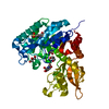
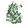
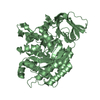
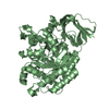
| Title | Structure of Chitinase D from Serratia proteamaculans revealed an unusually constrained substrate binding site |
|---|
 Components Components | Glycoside hydrolase family 18 |
|---|
 Keywords Keywords | HYDROLASE / TIM BARREL |
|---|
| Function / homology |  Function and homology information Function and homology information
endochitinase activity / chitinase / chitin catabolic process / chitin binding / polysaccharide catabolic processSimilarity search - Function Chitinase A; domain 3 - #10 / Glycosyl hydrolases family 18 (GH18) active site / Glycosyl hydrolases family 18 (GH18) active site signature. / : / Chitinase insertion domain superfamily / Chitinase II / Glyco_18 / Glycosyl hydrolases family 18 / Glycosyl hydrolases family 18 (GH18) domain profile. / Glycoside hydrolase family 18, catalytic domain ...Chitinase A; domain 3 - #10 / Glycosyl hydrolases family 18 (GH18) active site / Glycosyl hydrolases family 18 (GH18) active site signature. / : / Chitinase insertion domain superfamily / Chitinase II / Glyco_18 / Glycosyl hydrolases family 18 / Glycosyl hydrolases family 18 (GH18) domain profile. / Glycoside hydrolase family 18, catalytic domain / Chitinase A; domain 3 / Glycosidases / Glycoside hydrolase superfamily / TIM Barrel / Alpha-Beta Barrel / Roll / Alpha BetaSimilarity search - Domain/homology |
|---|
| Biological species |  Serratia proteamaculans (bacteria) Serratia proteamaculans (bacteria) |
|---|
| Method |  X-RAY DIFFRACTION / SYNCHROTRON / MOLECULAR REPLACEMENT / Resolution: 1.49 Å X-RAY DIFFRACTION / SYNCHROTRON / MOLECULAR REPLACEMENT / Resolution: 1.49 Å |
|---|
 Authors Authors | Madhuprakash, J. / Singh, A. / Kumar, S. / Sinha, M. / Kaur, P. / Sharma, S. / Podile, A.R. / Singh, T.P. |
|---|
 Citation Citation | Journal: Sci Rep / Year: 2015
Title: Inverse relationship between chitobiase and transglycosylation activities of chitinase-D from Serratia proteamaculans revealed by mutational and biophysical analyses.
Authors: Madhuprakash, J. / Bobbili, K.B. / Moerschbacher, B.M. / Singh, T.P. / Swamy, M.J. / Podile, A.R. |
|---|
| History | | Deposition | Jun 30, 2013 | Deposition site: RCSB / Processing site: PDBJ |
|---|
| Revision 1.0 | Oct 2, 2013 | Provider: repository / Type: Initial release |
|---|
| Revision 1.1 | Jul 27, 2016 | Group: Database references |
|---|
| Revision 1.2 | Nov 8, 2023 | Group: Data collection / Database references ...Data collection / Database references / Derived calculations / Refinement description
Category: chem_comp_atom / chem_comp_bond ...chem_comp_atom / chem_comp_bond / database_2 / pdbx_initial_refinement_model / struct_conn / struct_site
Item: _database_2.pdbx_DOI / _database_2.pdbx_database_accession ..._database_2.pdbx_DOI / _database_2.pdbx_database_accession / _struct_conn.pdbx_leaving_atom_flag / _struct_site.pdbx_auth_asym_id / _struct_site.pdbx_auth_comp_id / _struct_site.pdbx_auth_seq_id |
|---|
| Revision 1.3 | Nov 6, 2024 | Group: Structure summary / Category: pdbx_entry_details / pdbx_modification_feature |
|---|
|
|---|
 Movie
Movie Controller

 Yorodumi
 Open data
 Basic information
 Structure visualization
 Downloads & links
 Other downloads


 PDBj
PDBj Assembly
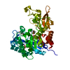

 Mass: 59.044 Da / Num. of mol.: 1 / Source method: obtained synthetically / Formula: C2H3O2
 Mass: 92.094 Da / Num. of mol.: 2 / Source method: obtained synthetically / Formula: C3H8O3
 Mass: 18.015 Da / Num. of mol.: 593 / Source method: isolated from a natural source / Formula: H2O
 Sample preparation
 / Beamline: BM14 / Wavelength: 0.97 Å
 Processing