 Movie
Movie Controller

[English] 日本語
 Yorodumi
- PDB-5hjl: Crystal structure of class I tagatose 1,6-bisphosphate aldolase L... -
+ Open data

- Basic information
| Entry | Database: PDB / ID: 5hjl | |||||||||
|---|---|---|---|---|---|---|---|---|---|---|
| Title | Crystal structure of class I tagatose 1,6-bisphosphate aldolase LacD from Streptococcus porcinus | |||||||||
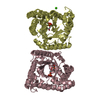
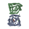
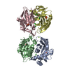
 Components Components | Tagatose 1,6-diphosphate aldolase | |||||||||
 Keywords Keywords | LYASE / tagatose 1 / 6-bisphosphate aldolase / LacD | |||||||||
| Function / homology | Aldolase class I / TIM Barrel / Alpha-Beta Barrel / Alpha Beta / :  Function and homology information Function and homology information | |||||||||
| Biological species |  Streptococcus porcinus str. Jelinkova 176 (bacteria) Streptococcus porcinus str. Jelinkova 176 (bacteria) | |||||||||
| Method |  X-RAY DIFFRACTION / SYNCHROTRON / MOLECULAR REPLACEMENT / molecular replacement / Resolution: 3 Å X-RAY DIFFRACTION / SYNCHROTRON / MOLECULAR REPLACEMENT / molecular replacement / Resolution: 3 Å | |||||||||
 Authors Authors | Freichels, R. / Kerff, F. / Herman, R. / Charlier, P. / Galleni, M. | |||||||||
| Funding support |  Belgium, 2items Belgium, 2items
| |||||||||
 Citation Citation | Journal: To Be Published Title: Structure and Characterization of a new class I Tagatose-1,6-bipshosphate aldolase from Streptococcus porcinus : switch in specificity directed by an Arginine Authors: Freichels, R. / Delmarcelle, M. / Van der Heiden, E. / Herman, R. / Colarusso, A. / Wathelet, B. / Kerff, F. / Galleni, M. | |||||||||
| History |
|
- Structure visualization
| Structure viewer | Molecule: MolmilJmol/JSmol |
|---|
- Downloads & links
-Download
| PDBx/mmCIF format | 5hjl.cif.gz | 134.8 KB | Display | PDBx/mmCIF format |
|---|---|---|---|---|
| PDB format | pdb5hjl.ent.gz | 105.5 KB | Display | PDB format |
| PDBx/mmJSON format | 5hjl.json.gz | Tree view | PDBx/mmJSON format | |
| Others |  Other downloads Other downloads |
-Validation report
| Arichive directory | https://data.pdbj.org/pub/pdb/validation_reports/hj/5hjlftp://data.pdbj.org/pub/pdb/validation_reports/hj/5hjl | HTTPS FTP |
|---|
-Related structure data
| Related structure data | 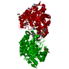 3iv3S S: Starting model for refinement |
|---|---|
| Similar structure data |


-Links
 PDBj
PDBj- Assembly
| Deposited unit | 
| |||||||||||||||||||||
|---|---|---|---|---|---|---|---|---|---|---|---|---|---|---|---|---|---|---|---|---|---|---|
| 1 |
| |||||||||||||||||||||
| Unit cell |
| |||||||||||||||||||||
| Noncrystallographic symmetry (NCS) | NCS domain:
NCS domain segments: Component-ID: 1 / Ens-ID: 1 / Beg auth comp-ID: SER / Beg label comp-ID: SER / End auth comp-ID: LEU / End label comp-ID: LEU / Auth seq-ID: 5 - 326 / Label seq-ID: 5 - 326
|
-Components
| #1: Protein | Mass: 36962.938 Da / Num. of mol.: 2 Source method: isolated from a genetically manipulated source Source: (gene. exp.) Streptococcus porcinus str. Jelinkova 176 (bacteria)Gene: lacD, STRPO_0644 / Plasmid: pET28a(+) / Production host: #2: Chemical | ChemComp-SO4 /   Mass: 96.063 Da / Num. of mol.: 6 / Source method: obtained synthetically / Formula: SO4 Mass: 96.063 Da / Num. of mol.: 6 / Source method: obtained synthetically / Formula: SO4#3: Water | ChemComp-HOH / |  Mass: 18.015 Da / Num. of mol.: 8 / Source method: isolated from a natural source / Formula: H2O Mass: 18.015 Da / Num. of mol.: 8 / Source method: isolated from a natural source / Formula: H2O |
|---|
-Experimental details
-Experiment
| Experiment | Method: X-RAY DIFFRACTION / Number of used crystals: 1 |
|---|
- Sample preparation
| Crystal | Density Matthews: 2.51 Å3/Da / Density % sol: 51.04 % |
|---|---|
| Crystal grow | Temperature: 293 K / Method: vapor diffusion, sitting drop / pH: 7.5 Details: Small crystals grow in 25% PEG 3350, 0.1M bistris pH 5.5, 0.2 M Ammonium Sulfate. The crystal used for data collection was obtained after micro seeding in the same condition. |
-Data collection
| Diffraction | Mean temperature: 100 K |
|---|---|
| Diffraction source | Source: SYNCHROTRON / Site: SOLEIL  / Beamline: PROXIMA 2 / Wavelength: 1.7101 Å / Beamline: PROXIMA 2 / Wavelength: 1.7101 Å |
| Detector | Type: DECTRIS PILATUS 6M / Detector: PIXEL / Date: Mar 21, 2015 |
| Radiation | Protocol: SINGLE WAVELENGTH / Monochromatic (M) / Laue (L): M / Scattering type: x-ray |
| Radiation wavelength | Wavelength: 1.7101 Å / Relative weight: 1 |
| Reflection | Resolution: 3→46.88 Å / Num. obs: 14194 / % possible obs: 99.9 % / Redundancy: 5.5 % / Rmerge(I) obs: 0.296 / Net I/σ(I): 5.9 |
| Reflection shell | Resolution: 3→3.16 Å / Redundancy: 5.6 % / Rmerge(I) obs: 1.13 / Mean I/σ(I) obs: 2.4 / % possible all: 100 |
-Phasing
| Phasing | Method: molecular replacement |
|---|
- Processing
| Software |
| ||||||||||||||||||||||||||||||||||||||||||
|---|---|---|---|---|---|---|---|---|---|---|---|---|---|---|---|---|---|---|---|---|---|---|---|---|---|---|---|---|---|---|---|---|---|---|---|---|---|---|---|---|---|---|---|
| Refinement | Method to determine structure: MOLECULAR REPLACEMENT Starting model: 3IV3 Resolution: 3→46.87 Å / SU ML: 0.39 / Cross valid method: FREE R-VALUE / Phase error: 25.01 / Stereochemistry target values: ML
| ||||||||||||||||||||||||||||||||||||||||||
| Solvent computation | Shrinkage radii: 0.9 Å / VDW probe radii: 1.11 Å / Solvent model: FLAT BULK SOLVENT MODEL | ||||||||||||||||||||||||||||||||||||||||||
| Displacement parameters | Biso max: 111.76 Å2 / Biso mean: 61.6768 Å2 / Biso min: 38.61 Å2 | ||||||||||||||||||||||||||||||||||||||||||
| Refinement step | Cycle: final / Resolution: 3→46.87 Å
| ||||||||||||||||||||||||||||||||||||||||||
| Refine LS restraints |
| ||||||||||||||||||||||||||||||||||||||||||
| Refine LS restraints NCS |
| ||||||||||||||||||||||||||||||||||||||||||
| LS refinement shell |
|
