 Movie
Movie Controller

[English] 日本語
 Yorodumi
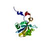
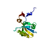
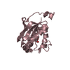
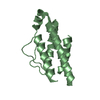
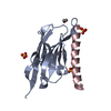
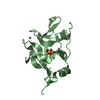
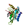
- PDB-5hfc: The third PDZ domain from the synaptic protein PSD-95 (H372A muta... -
+ Open data

- Basic information
| Entry | Database: PDB / ID: 5hfc | ||||||
|---|---|---|---|---|---|---|---|
| Title | The third PDZ domain from the synaptic protein PSD-95 (H372A mutant) in complex with a mutant C-terminal peptide derived from CRIPT (T-2F) | ||||||
 Components Components |
| ||||||
 Keywords Keywords | PEPTIDE BINDING PROTEIN / PDZ / GLGF / DHR / adhesion / synapse / synaptic density / peptide-binding domain | ||||||
| Function / homology |  Function and homology information Function and homology informationRHO GTPases activate CIT / regulation of postsynaptic density protein 95 clustering / neuronal ion channel clustering / positive regulation of AMPA glutamate receptor clustering / P2Y1 nucleotide receptor binding / Neurexins and neuroligins / beta-1 adrenergic receptor binding / neuroligin family protein binding / regulation of grooming behavior / structural constituent of postsynaptic density ...RHO GTPases activate CIT / regulation of postsynaptic density protein 95 clustering / neuronal ion channel clustering / positive regulation of AMPA glutamate receptor clustering / P2Y1 nucleotide receptor binding / Neurexins and neuroligins / beta-1 adrenergic receptor binding / neuroligin family protein binding / regulation of grooming behavior / structural constituent of postsynaptic density / synaptic vesicle maturation / protein localization to microtubule / positive regulation of neuron projection arborization / receptor localization to synapse / regulation of postsynaptic density assembly / cerebellar mossy fiber / LGI-ADAM interactions / protein localization to synapse / neuron spine / dendritic spine morphogenesis / Trafficking of AMPA receptors / proximal dendrite / negative regulation of receptor internalization / juxtaparanode region of axon / vocalization behavior / Activation of Ca-permeable Kainate Receptor / dendritic branch / acetylcholine receptor binding / positive regulation of dendrite morphogenesis / frizzled binding / cellular response to potassium ion / positive regulation of synapse assembly / dendritic spine organization / regulation of NMDA receptor activity / RAF/MAP kinase cascade / NMDA selective glutamate receptor signaling pathway / beta-2 adrenergic receptor binding / Synaptic adhesion-like molecules / neuromuscular process controlling balance / neurotransmitter receptor localization to postsynaptic specialization membrane / neuron projection terminus / cortical cytoskeleton / AMPA glutamate receptor clustering / locomotory exploration behavior / AMPA glutamate receptor complex / Unblocking of NMDA receptors, glutamate binding and activation / regulation of neuronal synaptic plasticity / glutamate receptor binding / social behavior / kinesin binding / D1 dopamine receptor binding / positive regulation of synaptic transmission / regulation of postsynaptic membrane neurotransmitter receptor levels / postsynaptic density, intracellular component / cytoplasmic microtubule organization / excitatory synapse / ionotropic glutamate receptor binding / dendrite cytoplasm / RNA splicing / positive regulation of excitatory postsynaptic potential / cell periphery / dendritic shaft / synaptic membrane / spliceosomal complex / PDZ domain binding / neuromuscular junction / establishment of protein localization / cell-cell adhesion / cerebral cortex development / regulation of long-term neuronal synaptic plasticity / postsynaptic density membrane / kinase binding / mRNA processing / cell-cell junction / synaptic vesicle / cell junction / nervous system development / positive regulation of cytosolic calcium ion concentration / protein-containing complex assembly / scaffold protein binding / protein phosphatase binding / microtubule binding / dendritic spine / chemical synaptic transmission / postsynaptic membrane / neuron projection / postsynaptic density / postsynapse / signaling receptor binding / neuronal cell body / synapse / dendrite / protein kinase binding / protein-containing complex binding / glutamatergic synapse / endoplasmic reticulum / membrane / plasma membrane / cytoplasm / cytosol Similarity search - Function | ||||||
| Biological species |  | ||||||
| Method |  X-RAY DIFFRACTION / MOLECULAR REPLACEMENT / Resolution: 1.851 Å X-RAY DIFFRACTION / MOLECULAR REPLACEMENT / Resolution: 1.851 Å | ||||||
 Authors Authors | White, K.I. / Raman, A.S. / Ranganathan, R. | ||||||
| Funding support |  United States, 1items United States, 1items
| ||||||
 Citation Citation | Journal: Cell(Cambridge,Mass.) / Year: 2016 Title: Origins of Allostery and Evolvability in Proteins: A Case Study. Authors: Raman, A.S. / White, K.I. / Ranganathan, R. | ||||||
| History |
|
- Structure visualization
| Structure viewer | Molecule: MolmilJmol/JSmol |
|---|
- Downloads & links
-Download
| PDBx/mmCIF format | 5hfc.cif.gz | 91 KB | Display | PDBx/mmCIF format |
|---|---|---|---|---|
| PDB format | pdb5hfc.ent.gz | 72.3 KB | Display | PDB format |
| PDBx/mmJSON format | 5hfc.json.gz | Tree view | PDBx/mmJSON format | |
| Others |  Other downloads Other downloads |
-Validation report
| Arichive directory | https://data.pdbj.org/pub/pdb/validation_reports/hf/5hfcftp://data.pdbj.org/pub/pdb/validation_reports/hf/5hfc | HTTPS FTP |
|---|
-Related structure data
| Related structure data |  5hebC  5hedC  5hetC 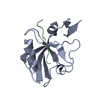 5heyC  5hf1C 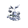 5hfbC  5hffC 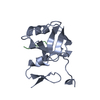 1be9S S: Starting model for refinement C: citing same article ( |
|---|---|
| Similar structure data |


-Links
 PDBj
PDBj


- Assembly
| Deposited unit | 
| |||||||||
|---|---|---|---|---|---|---|---|---|---|---|
| 1 |
| |||||||||
| Unit cell |
| |||||||||
| Components on special symmetry positions |
|
-Components
| #1: Protein | Mass: 12670.998 Da / Num. of mol.: 1 / Fragment: PDZ-3 domain (UNP residues 302-402) / Mutation: H372A Source method: isolated from a genetically manipulated source Source: (gene. exp.)  |
|---|---|
| #2: Protein/peptide | Mass: 1116.266 Da / Num. of mol.: 1 / Fragment: PDZ3-binding domain (UNP residues 93-101) / Mutation: T99F / Source method: obtained synthetically Details: Synthesized using standard FMOC chemistry, HPCL purified, and lyophilized. Source: (synth.) |
| #3: Water | ChemComp-HOH /  Mass: 18.015 Da / Num. of mol.: 124 / Source method: isolated from a natural source / Formula: H2O Mass: 18.015 Da / Num. of mol.: 124 / Source method: isolated from a natural source / Formula: H2O |
-Experimental details
-Experiment
| Experiment | Method: X-RAY DIFFRACTION / Number of used crystals: 1 |
|---|
- Sample preparation
| Crystal | Density Matthews: 2.15 Å3/Da / Density % sol: 43 % |
|---|---|
| Crystal grow | Temperature: 289 K / Method: vapor diffusion, hanging drop / pH: 7 Details: Peptide was included in protein buffer to a final molar ratio of 2:1 relative to protein. Reservoir solution contained 1.05 M sodium citrate, pH 7.0. Equal amounts (1.5 microliters) of ...Details: Peptide was included in protein buffer to a final molar ratio of 2:1 relative to protein. Reservoir solution contained 1.05 M sodium citrate, pH 7.0. Equal amounts (1.5 microliters) of protein (7 mg/mL) and reservoir solution were mixed and equillibrated against 500 microliters of crystallization buffer. |
-Data collection
| Diffraction | Mean temperature: 100 K |
|---|---|
| Diffraction source | Source: ROTATING ANODE / Type: RIGAKU FR-E SUPERBRIGHT / Wavelength: 1.54178 Å |
| Detector | Type: RIGAKU RAXIS IV++ / Detector: IMAGE PLATE / Date: Sep 11, 2012 |
| Radiation | Protocol: SINGLE WAVELENGTH / Monochromatic (M) / Laue (L): M / Scattering type: x-ray |
| Radiation wavelength | Wavelength: 1.54178 Å / Relative weight: 1 |
| Reflection | Resolution: 1.851→40.001 Å / Num. obs: 10760 / % possible obs: 98.22 % / Redundancy: 6.3 % / Rmerge(I) obs: 0.04 / Net I/σ(I): 35.543 |
| Reflection shell | Resolution: 1.8514→1.9357 Å / Redundancy: 2.5 % / Rmerge(I) obs: 0.448 / Mean I/σ(I) obs: 1 / % possible all: 88 |
- Processing
| Software |
| |||||||||||||||||||||||||||||||||||||||||||||||||||||||||||||||||||||||||||||||||||||||||||||||||||||||||||||||||||||||||||||||||||||||||||||||||||||||||||||||||||||||||||||||||||||||||||||||||||||||||||||||||||||||||||||||||
|---|---|---|---|---|---|---|---|---|---|---|---|---|---|---|---|---|---|---|---|---|---|---|---|---|---|---|---|---|---|---|---|---|---|---|---|---|---|---|---|---|---|---|---|---|---|---|---|---|---|---|---|---|---|---|---|---|---|---|---|---|---|---|---|---|---|---|---|---|---|---|---|---|---|---|---|---|---|---|---|---|---|---|---|---|---|---|---|---|---|---|---|---|---|---|---|---|---|---|---|---|---|---|---|---|---|---|---|---|---|---|---|---|---|---|---|---|---|---|---|---|---|---|---|---|---|---|---|---|---|---|---|---|---|---|---|---|---|---|---|---|---|---|---|---|---|---|---|---|---|---|---|---|---|---|---|---|---|---|---|---|---|---|---|---|---|---|---|---|---|---|---|---|---|---|---|---|---|---|---|---|---|---|---|---|---|---|---|---|---|---|---|---|---|---|---|---|---|---|---|---|---|---|---|---|---|---|---|---|---|---|---|---|---|---|---|---|---|---|---|---|---|---|---|---|---|---|
| Refinement | Method to determine structure: MOLECULAR REPLACEMENT Starting model: 1BE9 Resolution: 1.851→40.001 Å / SU ML: 0.18 / Cross valid method: FREE R-VALUE / σ(F): 1.46 / Phase error: 20.17 / Stereochemistry target values: ML
| |||||||||||||||||||||||||||||||||||||||||||||||||||||||||||||||||||||||||||||||||||||||||||||||||||||||||||||||||||||||||||||||||||||||||||||||||||||||||||||||||||||||||||||||||||||||||||||||||||||||||||||||||||||||||||||||||
| Solvent computation | Shrinkage radii: 0.9 Å / VDW probe radii: 1.11 Å / Solvent model: FLAT BULK SOLVENT MODEL | |||||||||||||||||||||||||||||||||||||||||||||||||||||||||||||||||||||||||||||||||||||||||||||||||||||||||||||||||||||||||||||||||||||||||||||||||||||||||||||||||||||||||||||||||||||||||||||||||||||||||||||||||||||||||||||||||
| Refinement step | Cycle: LAST / Resolution: 1.851→40.001 Å
| |||||||||||||||||||||||||||||||||||||||||||||||||||||||||||||||||||||||||||||||||||||||||||||||||||||||||||||||||||||||||||||||||||||||||||||||||||||||||||||||||||||||||||||||||||||||||||||||||||||||||||||||||||||||||||||||||
| Refine LS restraints |
| |||||||||||||||||||||||||||||||||||||||||||||||||||||||||||||||||||||||||||||||||||||||||||||||||||||||||||||||||||||||||||||||||||||||||||||||||||||||||||||||||||||||||||||||||||||||||||||||||||||||||||||||||||||||||||||||||
| LS refinement shell |
| |||||||||||||||||||||||||||||||||||||||||||||||||||||||||||||||||||||||||||||||||||||||||||||||||||||||||||||||||||||||||||||||||||||||||||||||||||||||||||||||||||||||||||||||||||||||||||||||||||||||||||||||||||||||||||||||||
| Refinement TLS params. | Method: refined / Refine-ID: X-RAY DIFFRACTION
| |||||||||||||||||||||||||||||||||||||||||||||||||||||||||||||||||||||||||||||||||||||||||||||||||||||||||||||||||||||||||||||||||||||||||||||||||||||||||||||||||||||||||||||||||||||||||||||||||||||||||||||||||||||||||||||||||
| Refinement TLS group |
|
