[English] 日本語
Yorodumi
- PDB-5hbc: Intermediate structure of iron-saturated C-lobe of bovine lactofe... -

+
Open data


ID or keywords:

Loading...

-
Basic information

Entry
Database: PDB / ID: 5hbc
Title
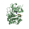
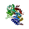
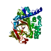
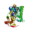
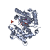
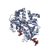
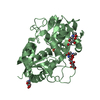
Intermediate structure of iron-saturated C-lobe of bovine lactoferrin at 2.79 Angstrom resolution indicates the softening of iron coordination
ComponentsLactotransferrinLactoferrin
KeywordsHYDROLASE / C-LOBE / Lactoferrin
Function / homology
Function and homology information


Metal sequestration by antimicrobial proteins / Antimicrobial peptides / negative regulation of cysteine-type endopeptidase activity / negative regulation of tumor necrosis factor (ligand) superfamily member 11 production / negative regulation of single-species biofilm formation in or on host organism / positive regulation of bone mineralization involved in bone maturation / negative regulation of lipopolysaccharide-mediated signaling pathway / specific granule / negative regulation of osteoclast development / antifungal humoral response ...Metal sequestration by antimicrobial proteins / Antimicrobial peptides / negative regulation of cysteine-type endopeptidase activity / negative regulation of tumor necrosis factor (ligand) superfamily member 11 production / negative regulation of single-species biofilm formation in or on host organism / positive regulation of bone mineralization involved in bone maturation / negative regulation of lipopolysaccharide-mediated signaling pathway / specific granule / negative regulation of osteoclast development / antifungal humoral response / positive regulation of chondrocyte proliferation / regulation of tumor necrosis factor production / bone morphogenesis / Neutrophil degranulation / cysteine-type endopeptidase inhibitor activity / positive regulation of osteoblast proliferation / Hydrolases; Acting on peptide bonds (peptidases); Serine endopeptidases / positive regulation of osteoblast differentiation / regulation of cytokine production / ossification / innate immune response in mucosa / recycling endosome / antibacterial humoral response / iron ion transport / early endosome / iron ion binding / signaling receptor binding / serine-type endopeptidase activity / negative regulation of apoptotic process / proteolysis / extracellular space / plasma membrane
Similarity search - Function
Lactotransferrin / Transferrin-like domain signature 2. / Transferrin family, iron binding site / Transferrin-like domain signature 1. / Transferrin-like domain signature 3. / Transferrin-like domain / Transferrin / Transferrin / Transferrin-like domain profile. / Transferrin ...Lactotransferrin / Transferrin-like domain signature 2. / Transferrin family, iron binding site / Transferrin-like domain signature 1. / Transferrin-like domain signature 3. / Transferrin-like domain / Transferrin / Transferrin / Transferrin-like domain profile. / Transferrin / Periplasmic binding protein-like II / D-Maltodextrin-Binding Protein; domain 2 / 3-Layer(aba) Sandwich / Alpha Beta
Similarity search - Domain/homology
BICARBONATE ION / : / Lactotransferrin
Similarity search - Component
Biological speciesBos taurus (cattle)
MethodX-RAY DIFFRACTION / SYNCHROTRON / MOLECULAR REPLACEMENT / Resolution: 2.79 Å
AuthorsSingh, A. / Rastogi, N. / Singh, P.K. / Tyagi, T.K. / Kaur, P. / Sharma, S. / Singh, T.P.
CitationJournal: Proteins / Year: 2016
Title: Structure of iron saturated C-lobe of bovine lactoferrin at pH 6.8 indicates a weakening of iron coordination
Authors: Rastogi, N. / Singh, A. / Singh, P.K. / Tyagi, T.K. / Pandey, S. / Shin, K. / Kaur, P. / Sharma, S. / Singh, T.P.
History
DepositionDec 31, 2015Deposition site: RCSB / Processing site: PDBJ
Revision 1.0Jan 20, 2016Provider: repository / Type: Initial release
Revision 1.1Jul 20, 2016Group: Database references
Revision 2.0Jul 29, 2020Group: Atomic model / Data collection ...Atomic model / Data collection / Database references / Derived calculations / Structure summary
Category: atom_site / chem_comp ...atom_site / chem_comp / citation / entity / pdbx_branch_scheme / pdbx_chem_comp_identifier / pdbx_entity_branch / pdbx_entity_branch_descriptor / pdbx_entity_branch_link / pdbx_entity_branch_list / pdbx_entity_nonpoly / pdbx_nonpoly_scheme / pdbx_struct_assembly_gen / pdbx_struct_conn_angle / pdbx_struct_oper_list / struct_asym / struct_conn / struct_site / struct_site_gen
Item: _atom_site.B_iso_or_equiv / _atom_site.Cartn_x ..._atom_site.B_iso_or_equiv / _atom_site.Cartn_x / _atom_site.Cartn_y / _atom_site.Cartn_z / _atom_site.auth_asym_id / _atom_site.auth_atom_id / _atom_site.auth_comp_id / _atom_site.auth_seq_id / _atom_site.label_asym_id / _atom_site.label_atom_id / _atom_site.label_comp_id / _atom_site.label_entity_id / _atom_site.type_symbol / _chem_comp.name / _chem_comp.type / _citation.journal_id_CSD / _entity.formula_weight / _entity.pdbx_description / _entity.pdbx_number_of_molecules / _entity.src_method / _entity.type / _pdbx_struct_assembly_gen.asym_id_list / _pdbx_struct_conn_angle.ptnr1_label_asym_id / _pdbx_struct_conn_angle.ptnr2_label_asym_id / _pdbx_struct_conn_angle.ptnr3_label_asym_id / _pdbx_struct_oper_list.symmetry_operation / _struct_conn.conn_type_id / _struct_conn.id / _struct_conn.pdbx_dist_value / _struct_conn.pdbx_leaving_atom_flag / _struct_conn.pdbx_role / _struct_conn.ptnr1_auth_asym_id / _struct_conn.ptnr1_auth_comp_id / _struct_conn.ptnr1_auth_seq_id / _struct_conn.ptnr1_label_asym_id / _struct_conn.ptnr1_label_atom_id / _struct_conn.ptnr1_label_comp_id / _struct_conn.ptnr1_label_seq_id / _struct_conn.ptnr2_auth_asym_id / _struct_conn.ptnr2_auth_comp_id / _struct_conn.ptnr2_auth_seq_id / _struct_conn.ptnr2_label_asym_id / _struct_conn.ptnr2_label_atom_id / _struct_conn.ptnr2_label_comp_id
Description: Carbohydrate remediation / Provider: repository / Type: Remediation
Revision 2.1Nov 8, 2023Group: Data collection / Database references ...Data collection / Database references / Refinement description / Structure summary
Category: chem_comp / chem_comp_atom ...chem_comp / chem_comp_atom / chem_comp_bond / database_2 / pdbx_initial_refinement_model
Item: _chem_comp.pdbx_synonyms / _database_2.pdbx_DOI / _database_2.pdbx_database_accession

-
Structure visualization

Structure viewerMolecule:
MolmilJmol/JSmol

Downloads & links

-
Assembly

Deposited unit
A: Lactotransferrin
B: Lactotransferrin
hetero molecules


Theoretical massNumber of molelcules
Total (without water)78,49011
Polymers76,0542
Non-polymers2,4369
Water1,27971
1
A: Lactotransferrin
hetero molecules


Theoretical massNumber of molelcules
Total (without water)39,3175
Polymers38,0271
Non-polymers1,2904
Water181
TypeNameSymmetry operationNumber
identity operation1_555x,y,z1
Buried area1730 Å2
ΔGint8 kcal/mol
Surface area15830 Å2
MethodPISA
2
B: Lactotransferrin
hetero molecules


Theoretical massNumber of molelcules
Total (without water)39,1736
Polymers38,0271
Non-polymers1,1465
Water181
TypeNameSymmetry operationNumber
identity operation1_555x,y,z1
Buried area1440 Å2
ΔGint8 kcal/mol
Surface area15850 Å2
MethodPISA
Unit cell
Length a, b, c (Å)159.654, 82.580, 107.694
Angle α, β, γ (deg.)90.00, 128.08, 90.00
Int Tables number5
Space group name H-MC121

-
Components

-
Protein , 1 types, 2 molecules AB

#1: Protein Lactotransferrin / Lactoferrin / Lactoferrin


Mass: 38026.961 Da / Num. of mol.: 2 / Fragment: UNP residues 361-708 / Source method: isolated from a natural source / Source: (natural) Bos taurus (cattle)
References: UniProt: P24627, Hydrolases; Acting on peptide bonds (peptidases); Serine endopeptidases

-
Sugars , 2 types, 5 molecules

#2: Polysaccharide beta-D-mannopyranose-(1-4)-2-acetamido-2-deoxy-beta-D-glucopyranose-(1-4)-2-acetamido-2-deoxy-beta- ...beta-D-mannopyranose-(1-4)-2-acetamido-2-deoxy-beta-D-glucopyranose-(1-4)-2-acetamido-2-deoxy-beta-D-glucopyranose


Type: oligosaccharide / Mass: 586.542 Da / Num. of mol.: 3
Source method: isolated from a genetically manipulated source
DescriptorTypeProgram
DManpb1-4DGlcpNAcb1-4DGlcpNAcb1-Glycam Condensed SequenceGMML 1.0
WURCS=2.0/2,3,2/[a2122h-1b_1-5_2*NCC/3=O][a1122h-1b_1-5]/1-1-2/a4-b1_b4-c1WURCSPDB2Glycan 1.1.0
[]{[(4+1)][b-D-GlcpNAc]{[(4+1)][b-D-GlcpNAc]{[(4+1)][b-D-Manp]{}}}}LINUCSPDB-CARE
#5: Sugar ChemComp-NAG / 2-acetamido-2-deoxy-beta-D-glucopyranose / N-acetyl-beta-D-glucosamine / 2-acetamido-2-deoxy-beta-D-glucose / 2-acetamido-2-deoxy-D-glucose / 2-acetamido-2-deoxy-glucose / N-ACETYL-D-GLUCOSAMINE / N-Acetylglucosamine


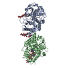
Type: D-saccharide, beta linking / Mass: 221.208 Da / Num. of mol.: 2
Source method: isolated from a genetically manipulated source
Formula: C8H15NO6
IdentifierTypeProgram
DGlcpNAcbCONDENSED IUPAC CARBOHYDRATE SYMBOLGMML 1.0
N-acetyl-b-D-glucopyranosamineCOMMON NAMEGMML 1.0
b-D-GlcpNAcIUPAC CARBOHYDRATE SYMBOLPDB-CARE 1.0
GlcNAcSNFG CARBOHYDRATE SYMBOLGMML 1.0

-
Non-polymers , 3 types, 75 molecules

#3: Chemical ChemComp-FE / FE (III) ION / Iron


Mass: 55.845 Da / Num. of mol.: 2 / Source method: obtained synthetically / Formula: Fe
#4: Chemical ChemComp-BCT / BICARBONATE ION / Bicarbonate


Mass: 61.017 Da / Num. of mol.: 2 / Source method: obtained synthetically / Formula: CHO3 / Comment: pH buffer*YM
#6: Water ChemComp-HOH / water / Water


Mass: 18.015 Da / Num. of mol.: 71 / Source method: isolated from a natural source / Formula: H2O

-
Details

Sequence detailsNATURAL MUTATIONS HAVE OCCURRED AT 584 ASN AND 627 LYS

-
Experimental details

-
Experiment

ExperimentMethod: X-RAY DIFFRACTION / Number of used crystals: 1

-
Sample preparation

CrystalDensity Matthews: 3.58 Å3/Da / Density % sol: 65.6 %
Crystal growTemperature: 300 K / Method: vapor diffusion, hanging drop / pH: 6.8 / Details: 0.05M HEPES, 20% PEG 20000

-
Data collection

DiffractionMean temperature: 80 K
Diffraction sourceSource: SYNCHROTRON / Site: ESRF / Beamline: BM14 / Wavelength: 0.97 Å
DetectorType: MARRESEARCH / Detector: CCD / Date: Dec 22, 2014
RadiationMonochromator: Graphite / Protocol: SINGLE WAVELENGTH / Monochromatic (M) / Laue (L): M / Scattering type: x-ray
Radiation wavelengthWavelength: 0.97 Å / Relative weight: 1
ReflectionResolution: 2.788→39.45 Å / Num. all: 27307 / Num. obs: 27307 / % possible obs: 99.2 % / Redundancy: 4.1 % / Net I/σ(I): 16
Reflection shellResolution: 2.788→2.86 Å

-
Processing

Software
NameVersionClassification
REFMAC5.7.0032refinement
HKL-2000data reduction
SCALEPACKdata scaling
PHASERphasing
RefinementMethod to determine structure: MOLECULAR REPLACEMENT
Starting model: 4NED
Resolution: 2.79→39.45 Å / Cor.coef. Fo:Fc: 0.941 / Cor.coef. Fo:Fc free: 0.895 / Cross valid method: THROUGHOUT / ESU R: 0.714 / ESU R Free: 0.359 / Stereochemistry target values: MAXIMUM LIKELIHOOD / Details: HYDROGENS HAVE BEEN ADDED IN THE RIDING POSITIONS
RfactorNum. reflection% reflectionSelection details
Rfree0.2727 1369 5 %RANDOM
Rwork0.2061 ---
obs0.20948 25919 98.47 %-
Solvent computationIon probe radii: 0.8 Å / Shrinkage radii: 0.8 Å / VDW probe radii: 1.2 Å / Solvent model: MASK
Displacement parametersBiso mean: 67.422 Å2
Baniso -1Baniso -2Baniso -3
1--0.27 Å2-0 Å20.18 Å2
2---3.15 Å2-0 Å2
3---1.47 Å2
Refinement stepCycle: 1 / Resolution: 2.79→39.45 Å
ProteinNucleic acidLigandSolventTotal
Num. atoms5316 0 155 71 5542
Refine LS restraints
Refine-IDTypeDev idealDev ideal targetNumber
X-RAY DIFFRACTIONr_bond_refined_d0.0180.0195608
X-RAY DIFFRACTIONr_bond_other_d00.025228
X-RAY DIFFRACTIONr_angle_refined_deg1.8571.9847661
X-RAY DIFFRACTIONr_angle_other_deg3.6833.00711991
X-RAY DIFFRACTIONr_dihedral_angle_1_deg7.3885694
X-RAY DIFFRACTIONr_dihedral_angle_2_deg40.20425.042238
X-RAY DIFFRACTIONr_dihedral_angle_3_deg20.11115916
X-RAY DIFFRACTIONr_dihedral_angle_4_deg21.8661526
X-RAY DIFFRACTIONr_chiral_restr0.1070.2874
X-RAY DIFFRACTIONr_gen_planes_refined0.0120.026318
X-RAY DIFFRACTIONr_gen_planes_other0.0250.021220
X-RAY DIFFRACTIONr_nbd_refined
X-RAY DIFFRACTIONr_nbd_other
X-RAY DIFFRACTIONr_nbtor_refined
X-RAY DIFFRACTIONr_nbtor_other
X-RAY DIFFRACTIONr_xyhbond_nbd_refined
X-RAY DIFFRACTIONr_xyhbond_nbd_other
X-RAY DIFFRACTIONr_metal_ion_refined
X-RAY DIFFRACTIONr_metal_ion_other
X-RAY DIFFRACTIONr_symmetry_vdw_refined
X-RAY DIFFRACTIONr_symmetry_vdw_other
X-RAY DIFFRACTIONr_symmetry_hbond_refined
X-RAY DIFFRACTIONr_symmetry_hbond_other
X-RAY DIFFRACTIONr_symmetry_metal_ion_refined
X-RAY DIFFRACTIONr_symmetry_metal_ion_other
X-RAY DIFFRACTIONr_mcbond_it4.8446.5352782
X-RAY DIFFRACTIONr_mcbond_other4.8446.5332781
X-RAY DIFFRACTIONr_mcangle_it7.4469.7923474
X-RAY DIFFRACTIONr_mcangle_other7.4459.7943475
X-RAY DIFFRACTIONr_scbond_it5.0657.0562826
X-RAY DIFFRACTIONr_scbond_other5.0327.062821
X-RAY DIFFRACTIONr_scangle_it
X-RAY DIFFRACTIONr_scangle_other7.62610.4094135
X-RAY DIFFRACTIONr_long_range_B_refined10.15552.7876509
X-RAY DIFFRACTIONr_long_range_B_other10.15352.7866507
X-RAY DIFFRACTIONr_rigid_bond_restr
X-RAY DIFFRACTIONr_sphericity_free
X-RAY DIFFRACTIONr_sphericity_bonded
LS refinement shellResolution: 2.788→2.86 Å / Total num. of bins used: 20
RfactorNum. reflection% reflection
Rfree0.435 77 -
Rwork0.347 1597 -
obs--82.18 %

+
About Yorodumi

-
News

-
Feb 9, 2022. New format data for meta-information of EMDB entries

New format data for meta-information of EMDB entries

  • Version 3 of the EMDB header file is now the official format.
  • The previous official version 1.9 will be removed from the archive.

Related info.:EMDB header

External links:wwPDB to switch to version 3 of the EMDB data model

-
Aug 12, 2020. Covid-19 info

Covid-19 info

URL: https://pdbj.org/emnavi/covid19.php

New page: Covid-19 featured information page in EM Navigator.

Related info.:Covid-19 info / Mar 5, 2020. Novel coronavirus structure data

+
Mar 5, 2020. Novel coronavirus structure data

Novel coronavirus structure data

Related info.:Yorodumi Speices / Aug 12, 2020. Covid-19 info

External links:COVID-19 featured content - PDBj / Molecule of the Month (242):Coronavirus Proteases

+
Jan 31, 2019. EMDB accession codes are about to change! (news from PDBe EMDB page)

EMDB accession codes are about to change! (news from PDBe EMDB page)

  • The allocation of 4 digits for EMDB accession codes will soon come to an end. Whilst these codes will remain in use, new EMDB accession codes will include an additional digit and will expand incrementally as the available range of codes is exhausted. The current 4-digit format prefixed with “EMD-” (i.e. EMD-XXXX) will advance to a 5-digit format (i.e. EMD-XXXXX), and so on. It is currently estimated that the 4-digit codes will be depleted around Spring 2019, at which point the 5-digit format will come into force.
  • The EM Navigator/Yorodumi systems omit the EMD- prefix.

Related info.:Q: What is EMD? / ID/Accession-code notation in Yorodumi/EM Navigator

External links:EMDB Accession Codes are Changing Soon! / Contact to PDBj

+
Jul 12, 2017. Major update of PDB

Major update of PDB

  • wwPDB released updated PDB data conforming to the new PDBx/mmCIF dictionary.
  • This is a major update changing the version number from 4 to 5, and with Remediation, in which all the entries are updated.
  • In this update, many items about electron microscopy experimental information are reorganized (e.g. em_software).
  • Now, EM Navigator and Yorodumi are based on the updated data.

External links:wwPDB Remediation / Enriched Model Files Conforming to OneDep Data Standards Now Available in the PDB FTP Archive

-
Yorodumi

Thousand views of thousand structures

  • Yorodumi is a browser for structure data from EMDB, PDB, SASBDB, etc.
  • This page is also the successor to EM Navigator detail page, and also detail information page/front-end page for Omokage search.
  • The word "yorodu" (or yorozu) is an old Japanese word meaning "ten thousand". "mi" (miru) is to see.

Related info.:EMDB / PDB / SASBDB / Comparison of 3 databanks / Yorodumi Search / Aug 31, 2016. New EM Navigator & Yorodumi / Yorodumi Papers / Jmol/JSmol / Function and homology information / Changes in new EM Navigator and Yorodumi

Read more