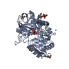
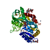
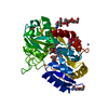
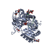
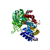
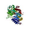
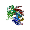
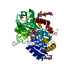
- PDB-4ned: Crystal STRUCTURE OF C-LOBE OF BOVINE LACTOFERRIN COMPLEXED WITH ... -
+
Open data
ID or keywords:
Loading...
-
Basic information
Entry
Database: PDB / ID: 4ned
Title
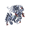
Crystal STRUCTURE OF C-LOBE OF BOVINE LACTOFERRIN COMPLEXED WITH FENOPROFEN AT 2.1 ANGSTROM RESOLUTION
Components
Lactotransferrin
Keywords
HYDROLASE / C-LOBE / Fenoprofen
Function / homology
Function and homology information
Metal sequestration by antimicrobial proteins / Antimicrobial peptides / negative regulation of tumor necrosis factor (ligand) superfamily member 11 production / negative regulation of single-species biofilm formation in or on host organism / positive regulation of bone mineralization involved in bone maturation / negative regulation of osteoclast development / specific granule / negative regulation of lipopolysaccharide-mediated signaling pathway / positive regulation of chondrocyte proliferation / antifungal humoral response ...Metal sequestration by antimicrobial proteins / Antimicrobial peptides / negative regulation of tumor necrosis factor (ligand) superfamily member 11 production / negative regulation of single-species biofilm formation in or on host organism / positive regulation of bone mineralization involved in bone maturation / negative regulation of osteoclast development / specific granule / negative regulation of lipopolysaccharide-mediated signaling pathway / positive regulation of chondrocyte proliferation / antifungal humoral response / regulation of tumor necrosis factor production / bone morphogenesis / Neutrophil degranulation / positive regulation of osteoblast proliferation / Hydrolases; Acting on peptide bonds (peptidases); Serine endopeptidases / positive regulation of osteoblast differentiation / cysteine-type endopeptidase inhibitor activity / regulation of cytokine production / ossification / innate immune response in mucosa / iron ion transport / recycling endosome / antibacterial humoral response / early endosome / iron ion binding / signaling receptor binding / serine-type endopeptidase activity / negative regulation of apoptotic process / proteolysis / : / plasma membrane Similarity search - Function
Mass: 18.015 Da / Num. of mol.: 204 / Source method: isolated from a natural source / Formula: H2O
-
Details
Has protein modification
Y
Sequence details
NATURAL MUTATIONS HAVE OCCURRED AT 584ASN AND 627LYS. AT PRESENT THESE MUTATIONS ARE NOT REPORTED ...NATURAL MUTATIONS HAVE OCCURRED AT 584ASN AND 627LYS. AT PRESENT THESE MUTATIONS ARE NOT REPORTED IN UNIPROT KB.
-
Experimental details
-
Experiment
Experiment
Method: X-RAY DIFFRACTION / Number of used crystals: 1
-
Sample preparation
Crystal
Density Matthews: 2.6 Å3/Da / Density % sol: 52.63 % Description: THE ENTRY CONTAINS FRIEDEL PAIRS IN F_PLUS/MINUS COLUMNS.
In the structure databanks used in Yorodumi, some data are registered as the other names, "COVID-19 virus" and "2019-nCoV". Here are the details of the virus and the list of structure data.
Jan 31, 2019. EMDB accession codes are about to change! (news from PDBe EMDB page)
EMDB accession codes are about to change! (news from PDBe EMDB page)
The allocation of 4 digits for EMDB accession codes will soon come to an end. Whilst these codes will remain in use, new EMDB accession codes will include an additional digit and will expand incrementally as the available range of codes is exhausted. The current 4-digit format prefixed with “EMD-” (i.e. EMD-XXXX) will advance to a 5-digit format (i.e. EMD-XXXXX), and so on. It is currently estimated that the 4-digit codes will be depleted around Spring 2019, at which point the 5-digit format will come into force.
The EM Navigator/Yorodumi systems omit the EMD- prefix.
Related info.:Q: What is EMD? / ID/Accession-code notation in Yorodumi/EM Navigator
Yorodumi is a browser for structure data from EMDB, PDB, SASBDB, etc.
This page is also the successor to EM Navigator detail page, and also detail information page/front-end page for Omokage search.
The word "yorodu" (or yorozu) is an old Japanese word meaning "ten thousand". "mi" (miru) is to see.
Related info.:EMDB / PDB / SASBDB / Comparison of 3 databanks / Yorodumi Search / Aug 31, 2016. New EM Navigator & Yorodumi / Yorodumi Papers / Jmol/JSmol / Function and homology information / Changes in new EM Navigator and Yorodumi
 Movie
Movie Controller

 Yorodumi
 Open data
 Basic information
 Components
 Keywords
 Function and homology information
 X-RAY DIFFRACTION /
  Authors
 Citation
 Structure visualization
 Downloads & links
 Other downloads

 PDBj
PDBj


 Assembly
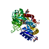

 Type: D-saccharide, beta linking / Mass: 221.208 Da / Num. of mol.: 2


 Mass: 65.409 Da / Num. of mol.: 2 / Source method: obtained synthetically / Formula: Zn
 Mass: 55.845 Da / Num. of mol.: 1 / Source method: obtained synthetically / Formula: Fe
 Mass: 60.009 Da / Num. of mol.: 1 / Source method: obtained synthetically / Formula: CO3
 Mass: 96.063 Da / Num. of mol.: 1 / Source method: obtained synthetically / Formula: SO4
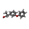
 Mass: 242.270 Da / Num. of mol.: 1 / Source method: obtained synthetically / Formula: C15H14O3
 Sample preparation
 / Beamline: BM14 / Wavelength: 0.97
  Processing