 Movie
Movie Controller

[English] 日本語
 Yorodumi
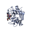
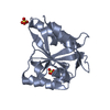
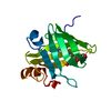
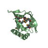
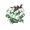
- PDB-5hae: Crystal structure of LTBP1 LTC4 complex collected on an in-house ... -
+ Open data

- Basic information
| Entry | Database: PDB / ID: 5hae | ||||||
|---|---|---|---|---|---|---|---|
| Title | Crystal structure of LTBP1 LTC4 complex collected on an in-house source | ||||||
 Components Components | Lipocalin AI-4 | ||||||
 Keywords Keywords | TRANSPORT PROTEIN / lipocalin / salivary / Rhodnius | ||||||
| Function / homology |  Function and homology information Function and homology information | ||||||
| Biological species |  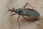 Rhodnius prolixus (insect) Rhodnius prolixus (insect) | ||||||
| Method |  X-RAY DIFFRACTION / FOURIER SYNTHESIS / Resolution: 2.002 Å X-RAY DIFFRACTION / FOURIER SYNTHESIS / Resolution: 2.002 Å | ||||||
 Authors Authors | Andersen, J.F. | ||||||
 Citation Citation | Journal: Acs Chem.Biol. / Year: 2016 Title: Structure and Ligand-Binding Mechanism of a Cysteinyl Leukotriene-Binding Protein from a Blood-Feeding Disease Vector. Authors: Jablonka, W. / Pham, V. / Nardone, G. / Gittis, A. / Silva-Cardoso, L. / Atella, G.C. / Ribeiro, J.M. / Andersen, J.F. | ||||||
| History |
|
- Structure visualization
| Structure viewer | Molecule: MolmilJmol/JSmol |
|---|
- Downloads & links
-Download
| PDBx/mmCIF format | 5hae.cif.gz | 84.8 KB | Display | PDBx/mmCIF format |
|---|---|---|---|---|
| PDB format | pdb5hae.ent.gz | 61.3 KB | Display | PDB format |
| PDBx/mmJSON format | 5hae.json.gz | Tree view | PDBx/mmJSON format | |
| Others |  Other downloads Other downloads |
-Validation report
| Arichive directory | https://data.pdbj.org/pub/pdb/validation_reports/ha/5haeftp://data.pdbj.org/pub/pdb/validation_reports/ha/5hae | HTTPS FTP |
|---|
-Related structure data


-Links
 PDBj
PDBj


- Assembly
| Deposited unit | 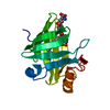
| ||||||||
|---|---|---|---|---|---|---|---|---|---|
| 1 |
| ||||||||
| Unit cell |
|
-Components
| #1: Protein | Mass: 16904.709 Da / Num. of mol.: 1 Source method: isolated from a genetically manipulated source Source: (gene. exp.) Rhodnius prolixus (insect) / Plasmid: pET17b / Production host:  |
|---|---|
| #2: Chemical | ChemComp-EAH / (  Mass: 320.466 Da / Num. of mol.: 1 / Source method: obtained synthetically / Formula: C20H32O3 Mass: 320.466 Da / Num. of mol.: 1 / Source method: obtained synthetically / Formula: C20H32O3 |
| #3: Chemical | ChemComp-GSH /   Mass: 307.323 Da / Num. of mol.: 1 / Source method: obtained synthetically / Formula: C10H17N3O6S Mass: 307.323 Da / Num. of mol.: 1 / Source method: obtained synthetically / Formula: C10H17N3O6S |
| #4: Water | ChemComp-HOH /  Mass: 18.015 Da / Num. of mol.: 168 / Source method: isolated from a natural source / Formula: H2O Mass: 18.015 Da / Num. of mol.: 168 / Source method: isolated from a natural source / Formula: H2O |
| Has protein modification | Y |
-Experimental details
-Experiment
| Experiment | Method: X-RAY DIFFRACTION / Number of used crystals: 1 |
|---|
- Sample preparation
| Crystal | Density Matthews: 2.07 Å3/Da / Density % sol: 40.59 % |
|---|---|
| Crystal grow | Temperature: 298 K / Method: vapor diffusion, hanging drop / pH: 8 / Details: 0.1M Tris, pH 8, 30% PEG 6000 / PH range: 7-8 |
-Data collection
| Diffraction | Mean temperature: 100 K | |||||||||||||||||||||||||||||||||||||||||||||||||||||||||||||||||||||||||||||||||||||||||||||||||||||||||||||||||||||||||||||||||||||||||||||||||||||||||||||||||||||||||||||||||||||||||||||
|---|---|---|---|---|---|---|---|---|---|---|---|---|---|---|---|---|---|---|---|---|---|---|---|---|---|---|---|---|---|---|---|---|---|---|---|---|---|---|---|---|---|---|---|---|---|---|---|---|---|---|---|---|---|---|---|---|---|---|---|---|---|---|---|---|---|---|---|---|---|---|---|---|---|---|---|---|---|---|---|---|---|---|---|---|---|---|---|---|---|---|---|---|---|---|---|---|---|---|---|---|---|---|---|---|---|---|---|---|---|---|---|---|---|---|---|---|---|---|---|---|---|---|---|---|---|---|---|---|---|---|---|---|---|---|---|---|---|---|---|---|---|---|---|---|---|---|---|---|---|---|---|---|---|---|---|---|---|---|---|---|---|---|---|---|---|---|---|---|---|---|---|---|---|---|---|---|---|---|---|---|---|---|---|---|---|---|---|---|---|---|
| Diffraction source | Source: ROTATING ANODE / Type: RIGAKU FR-E SUPERBRIGHT / Wavelength: 1.5418 Å | |||||||||||||||||||||||||||||||||||||||||||||||||||||||||||||||||||||||||||||||||||||||||||||||||||||||||||||||||||||||||||||||||||||||||||||||||||||||||||||||||||||||||||||||||||||||||||||
| Detector | Type: RIGAKU RAXIS IV / Detector: IMAGE PLATE / Date: Oct 9, 2015 | |||||||||||||||||||||||||||||||||||||||||||||||||||||||||||||||||||||||||||||||||||||||||||||||||||||||||||||||||||||||||||||||||||||||||||||||||||||||||||||||||||||||||||||||||||||||||||||
| Radiation | Protocol: SINGLE WAVELENGTH / Monochromatic (M) / Laue (L): M / Scattering type: x-ray | |||||||||||||||||||||||||||||||||||||||||||||||||||||||||||||||||||||||||||||||||||||||||||||||||||||||||||||||||||||||||||||||||||||||||||||||||||||||||||||||||||||||||||||||||||||||||||||
| Radiation wavelength | Wavelength: 1.5418 Å / Relative weight: 1 | |||||||||||||||||||||||||||||||||||||||||||||||||||||||||||||||||||||||||||||||||||||||||||||||||||||||||||||||||||||||||||||||||||||||||||||||||||||||||||||||||||||||||||||||||||||||||||||
| Reflection | Resolution: 2→50 Å / Num. obs: 9042 / % possible obs: 96.4 % / Redundancy: 3.9 % / Biso Wilson estimate: 16.31 Å2 / Rmerge(I) obs: 0.064 / Rpim(I) all: 0.036 / Rrim(I) all: 0.074 / Χ2: 2.666 / Net I/av σ(I): 37.619 / Net I/σ(I): 20.6 / Num. measured all: 35561 | |||||||||||||||||||||||||||||||||||||||||||||||||||||||||||||||||||||||||||||||||||||||||||||||||||||||||||||||||||||||||||||||||||||||||||||||||||||||||||||||||||||||||||||||||||||||||||||
| Reflection shell | Diffraction-ID: 1 / Rejects: _
|
- Processing
| Software |
| ||||||||||||||||||||||||||||||||||||||||
|---|---|---|---|---|---|---|---|---|---|---|---|---|---|---|---|---|---|---|---|---|---|---|---|---|---|---|---|---|---|---|---|---|---|---|---|---|---|---|---|---|---|
| Refinement | Method to determine structure: FOURIER SYNTHESIS / Resolution: 2.002→34.986 Å / SU ML: 0.2 / Cross valid method: FREE R-VALUE / σ(F): 1.4 / Phase error: 21.44 / Stereochemistry target values: ML
| ||||||||||||||||||||||||||||||||||||||||
| Solvent computation | Shrinkage radii: 0.9 Å / VDW probe radii: 1.11 Å / Solvent model: FLAT BULK SOLVENT MODEL | ||||||||||||||||||||||||||||||||||||||||
| Displacement parameters | Biso max: 79.63 Å2 / Biso mean: 20.6966 Å2 / Biso min: 7.78 Å2 | ||||||||||||||||||||||||||||||||||||||||
| Refinement step | Cycle: final / Resolution: 2.002→34.986 Å
| ||||||||||||||||||||||||||||||||||||||||
| Refine LS restraints |
| ||||||||||||||||||||||||||||||||||||||||
| LS refinement shell | Refine-ID: X-RAY DIFFRACTION / Total num. of bins used: 3
| ||||||||||||||||||||||||||||||||||||||||
| Refinement TLS params. | Method: refined / Origin x: -6.1287 Å / Origin y: 5.8651 Å / Origin z: -9.9589 Å
| ||||||||||||||||||||||||||||||||||||||||
| Refinement TLS group |
|
