| Entry | Database: PDB / ID: 5h7v
|
|---|
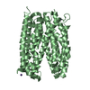
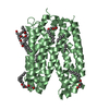
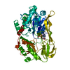
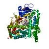
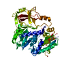
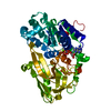
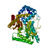
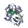
| Title | Structure of full-length extracellular domain of HAI-1 at pH 4.6 |
|---|
 Components Components | Kunitz-type protease inhibitor 1 |
|---|
 Keywords Keywords | HYDROLASE INHIBITOR / HAI-1 |
|---|
| Function / homology |  Function and homology information Function and homology information
epithelium development / Signaling by MST1 / positive regulation of glial cell differentiation / negative regulation of neural precursor cell proliferation / MET Receptor Activation / branching involved in labyrinthine layer morphogenesis / cellular response to BMP stimulus / placenta blood vessel development / epidermis development / extracellular matrix organization ...epithelium development / Signaling by MST1 / positive regulation of glial cell differentiation / negative regulation of neural precursor cell proliferation / MET Receptor Activation / branching involved in labyrinthine layer morphogenesis / cellular response to BMP stimulus / placenta blood vessel development / epidermis development / extracellular matrix organization / serine-type endopeptidase inhibitor activity / neural tube closure / : / extracellular exosome / extracellular region / membrane / plasma membrane / cytoplasmSimilarity search - Function MANEC domain / Seven cysteines, N-terminal / MANEC / MANSC domain / MANSC domain profile. / K319L-like, PKD domain / Pancreatic trypsin inhibitor Kunitz domain / Factor Xa Inhibitor / Low-density lipoprotein receptor domain class A / Low-density lipoprotein (LDL) receptor class A, conserved site ...MANEC domain / Seven cysteines, N-terminal / MANEC / MANSC domain / MANSC domain profile. / K319L-like, PKD domain / Pancreatic trypsin inhibitor Kunitz domain / Factor Xa Inhibitor / Low-density lipoprotein receptor domain class A / Low-density lipoprotein (LDL) receptor class A, conserved site / LDL-receptor class A (LDLRA) domain signature. / LDL-receptor class A (LDLRA) domain profile. / Low-density lipoprotein receptor domain class A / Low-density lipoprotein (LDL) receptor class A repeat / LDL receptor-like superfamily / Proteinase inhibitor I2, Kunitz, conserved site / Pancreatic trypsin inhibitor (Kunitz) family signature. / BPTI/Kunitz family of serine protease inhibitors. / Pancreatic trypsin inhibitor Kunitz domain / Kunitz/Bovine pancreatic trypsin inhibitor domain / Pancreatic trypsin inhibitor (Kunitz) family profile. / Pancreatic trypsin inhibitor Kunitz domain superfamily / Few Secondary Structures / Irregular / Immunoglobulin-like foldSimilarity search - Domain/homology |
|---|
| Biological species |  Homo sapiens (human) Homo sapiens (human) |
|---|
| Method |  X-RAY DIFFRACTION / SYNCHROTRON / MOLECULAR REPLACEMENT / Resolution: 3.82 Å X-RAY DIFFRACTION / SYNCHROTRON / MOLECULAR REPLACEMENT / Resolution: 3.82 Å |
|---|
 Authors Authors | Liu, M. / Huang, M. |
|---|
 Citation Citation | Journal: J. Biol. Chem. / Year: 2017
Title: The crystal structure of a multidomain protease inhibitor (HAI-1) reveals the mechanism of its auto-inhibition
Authors: Liu, M. / Yuan, C. / Jensen, J.K. / Zhao, B. / Jiang, Y. / Jiang, L. / Huang, M. |
|---|
| History | | Deposition | Nov 21, 2016 | Deposition site: PDBJ / Processing site: PDBJ |
|---|
| Revision 1.0 | Mar 29, 2017 | Provider: repository / Type: Initial release |
|---|
| Revision 1.1 | Apr 19, 2017 | Group: Database references |
|---|
| Revision 1.2 | May 31, 2017 | Group: Database references |
|---|
| Revision 1.3 | Nov 8, 2023 | Group: Data collection / Database references / Refinement description
Category: chem_comp_atom / chem_comp_bond ...chem_comp_atom / chem_comp_bond / database_2 / pdbx_initial_refinement_model
Item: _database_2.pdbx_DOI / _database_2.pdbx_database_accession |
|---|
| Revision 1.4 | Oct 23, 2024 | Group: Structure summary / Category: pdbx_entry_details / pdbx_modification_feature |
|---|
|
|---|
 Movie
Movie Controller


 Open data
 Basic information
 Structure visualization
 Downloads & links
 Other downloads
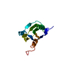


 PDBj
PDBj


 Assembly
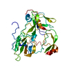

 Sample preparation
 / Beamline: BL18U1 / Wavelength: 0.9778 Å
 Processing