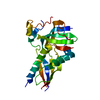
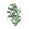
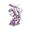
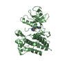
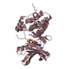
Mitochondrial distribution and morphology protein 12
Keywords
LIPID BINDING PROTEIN
Function / homology
Function and homology information
ERMES complex / protein insertion into mitochondrial outer membrane / phospholipid transport / : / lipid binding / endoplasmic reticulum membrane Similarity search - Function
Maintenance of mitochondrial morphology protein 1 / Mitochondrial distribution and morphology protein 12 / Mitochondrial distribution and morphology protein 12 / Synaptotagmin-like mitochondrial-lipid-binding domain / Synaptotagmin-like mitochondrial lipid-binding proteins (SMP) domain profile. Similarity search - Domain/homology
Protocol: SINGLE WAVELENGTH / Monochromatic (M) / Laue (L): M / Scattering type: x-ray
Radiation wavelength
Wavelength: 1 Å / Relative weight: 1
Reflection
Resolution: 3.31→50 Å / Num. obs: 535031 / % possible obs: 99.6 % / Redundancy: 10.7 % / Net I/σ(I): 15.6
-
Processing
Software
Name
Version
Classification
REFMAC
5.8.0073
refinement
HKL-2000
datareduction
HKL-2000
datascaling
MOLREP
phasing
Refinement
Method to determine structure: MOLECULAR REPLACEMENT / Resolution: 3.5→39.7 Å / Cor.coef. Fo:Fc: 0.894 / Cor.coef. Fo:Fc free: 0.849 / SU B: 30.762 / SU ML: 0.477 / Cross valid method: THROUGHOUT / ESU R Free: 0.621 / Details: HYDROGENS HAVE BEEN ADDED IN THE RIDING POSITIONS
Rfactor
Num. reflection
% reflection
Selection details
Rfree
0.32312
260
4.5 %
RANDOM
Rwork
0.26017
-
-
-
obs
0.26301
5467
99.81 %
-
Solvent computation
Ion probe radii: 0.8 Å / Shrinkage radii: 0.8 Å / VDW probe radii: 1.2 Å
 Movie
Movie Controller


 Open data
 Basic information
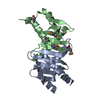
 Components
 Keywords
 Function and homology information
 Kluyveromyces lactis (yeast)
 X-RAY DIFFRACTION /
  Authors
 Japan, 1items
  Citation
 Structure visualization
 Downloads & links
 Other downloads
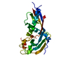


 PDBj
PDBj Assembly
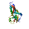

 Sample preparation
 Processing