 Movie
Movie Controller


+ Open data

- Basic information
| Entry | Database: PDB / ID: 5h0k | |||||||||||||||
|---|---|---|---|---|---|---|---|---|---|---|---|---|---|---|---|---|
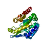
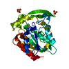
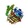
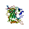
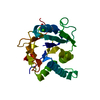
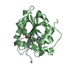
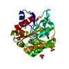
| Title | The crystal structure of WT Pedobacter heparinus SMUG2 | |||||||||||||||
 Components Components | Uncharacterized protein | |||||||||||||||
 Keywords Keywords | LYASE / SMUG / DNA damage / glycosylase | |||||||||||||||
| Function / homology | DNA glycosylase Phe SMUG2-like / : / Uracil-DNA glycosylase-like / Uracil DNA glycosylase superfamily / Uracil-DNA glycosylase-like domain superfamily / Uracil-DNA glycosylase-like domain-containing protein Function and homology information Function and homology information | |||||||||||||||
| Biological species |  Pedobacter heparinus DSM 2366 (bacteria) Pedobacter heparinus DSM 2366 (bacteria) | |||||||||||||||
| Method |  X-RAY DIFFRACTION / SYNCHROTRON / MOLECULAR REPLACEMENT / Resolution: 2.25 Å X-RAY DIFFRACTION / SYNCHROTRON / MOLECULAR REPLACEMENT / Resolution: 2.25 Å | |||||||||||||||
 Authors Authors | Xie, W. / Cao, W. / Pang, P. | |||||||||||||||
| Funding support |  China, 4items China, 4items
| |||||||||||||||
 Citation Citation | Journal: Biochem. J. / Year: 2017 Title: SMUG2 DNA glycosylase from Pedobacter heparinus as a new subfamily of the UDG superfamily Authors: Pang, P. / Yang, Y. / Li, J. / Wang, Z. / Cao, W. / Xie, W. | |||||||||||||||
| History |
|
- Structure visualization
| Structure viewer | Molecule: MolmilJmol/JSmol |
|---|
- Downloads & links
-Download
| PDBx/mmCIF format | 5h0k.cif.gz | 63.2 KB | Display | PDBx/mmCIF format |
|---|---|---|---|---|
| PDB format | pdb5h0k.ent.gz | 45 KB | Display | PDB format |
| PDBx/mmJSON format | 5h0k.json.gz | Tree view | PDBx/mmJSON format | |
| Others |  Other downloads Other downloads |
-Validation report
| Arichive directory | https://data.pdbj.org/pub/pdb/validation_reports/h0/5h0kftp://data.pdbj.org/pub/pdb/validation_reports/h0/5h0k | HTTPS FTP |
|---|
-Related structure data


-Links
 PDBj
PDBj- Assembly
| Deposited unit | 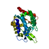
| |||||||||
|---|---|---|---|---|---|---|---|---|---|---|
| 1 |
| |||||||||
| Unit cell |
| |||||||||
| Components on special symmetry positions |
|
-Components
| #1: Protein | Mass: 28314.516 Da / Num. of mol.: 1 / Mutation: G65Y Source method: isolated from a genetically manipulated source Source: (gene. exp.) Pedobacter heparinus DSM 2366 (bacteria)Strain: DSM 2366 / Production host: |
|---|---|
| #2: Water | ChemComp-HOH /  Mass: 18.015 Da / Num. of mol.: 147 / Source method: isolated from a natural source / Formula: H2O Mass: 18.015 Da / Num. of mol.: 147 / Source method: isolated from a natural source / Formula: H2O |
-Experimental details
-Experiment
| Experiment | Method: X-RAY DIFFRACTION / Number of used crystals: 1 |
|---|
- Sample preparation
| Crystal | Density Matthews: 2.05 Å3/Da / Density % sol: 40.07 % |
|---|---|
| Crystal grow | Temperature: 298 K / Method: vapor diffusion, sitting drop / pH: 8 Details: 27% PEG 3350,0.25 M NaCl, 0.1M Tris-HCl pH 8.5/0.1M Hepes pH 8.0 |
-Data collection
| Diffraction | Mean temperature: 100 K |
|---|---|
| Diffraction source | Source: SYNCHROTRON / Site: SSRF / Beamline: BL17U / Wavelength: 0.979 Å |
| Detector | Type: ADSC QUANTUM 315 / Detector: CCD / Date: Apr 2, 2016 |
| Radiation | Protocol: SINGLE WAVELENGTH / Monochromatic (M) / Laue (L): M / Scattering type: x-ray |
| Radiation wavelength | Wavelength: 0.979 Å / Relative weight: 1 |
| Reflection | Resolution: 2.25→50 Å / Num. obs: 12005 / % possible obs: 99.9 % / Redundancy: 8.6 % / Rmerge(I) obs: 0.161 / Net I/σ(I): 19.7 |
| Reflection shell | Resolution: 2.25→2.33 Å / Redundancy: 8.7 % / Rmerge(I) obs: 0.639 / Mean I/σ(I) obs: 8.5 / CC1/2: 0.946 / % possible all: 99.8 |
- Processing
| Software |
| |||||||||||||||||||||||||||||||||||
|---|---|---|---|---|---|---|---|---|---|---|---|---|---|---|---|---|---|---|---|---|---|---|---|---|---|---|---|---|---|---|---|---|---|---|---|---|
| Refinement | Method to determine structure: MOLECULAR REPLACEMENT / Resolution: 2.25→38.643 Å / SU ML: 0.22 / Cross valid method: FREE R-VALUE / σ(F): 1.34 / Phase error: 25.52
| |||||||||||||||||||||||||||||||||||
| Solvent computation | Shrinkage radii: 0.9 Å / VDW probe radii: 1.11 Å | |||||||||||||||||||||||||||||||||||
| Refinement step | Cycle: LAST / Resolution: 2.25→38.643 Å
| |||||||||||||||||||||||||||||||||||
| Refine LS restraints |
| |||||||||||||||||||||||||||||||||||
| LS refinement shell |
|
