 Movie
Movie Controller

[English] 日本語
 Yorodumi
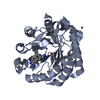
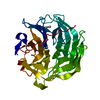
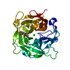
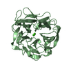
- PDB-5gx5: Luciferin-regenerating enzyme collected with serial synchrotron r... -
+ Open data

- Basic information
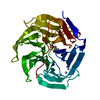
| Entry | Database: PDB / ID: 5gx5 | ||||||
|---|---|---|---|---|---|---|---|
| Title | Luciferin-regenerating enzyme collected with serial synchrotron rotational crystallography with accumulated dose of 26 MGy (23rd measurement) | ||||||
 Components Components | Luciferin regenerating enzyme | ||||||
 Keywords Keywords | HYDROLASE / BETA-PROOELLER | ||||||
| Function / homology |  Function and homology information Function and homology informationgluconolactonase / gluconolactonase activity / L-ascorbic acid biosynthetic process / calcium ion binding / cytoplasm Similarity search - Function | ||||||
| Biological species |  Photinus pyralis (common eastern firefly) Photinus pyralis (common eastern firefly) | ||||||
| Method |  X-RAY DIFFRACTION / SYNCHROTRON / FOURIER SYNTHESIS / Resolution: 1.6 Å X-RAY DIFFRACTION / SYNCHROTRON / FOURIER SYNTHESIS / Resolution: 1.6 Å | ||||||
 Authors Authors | Hasegawa, K. / Yamashita, K. / Murai, T. / Nuemket, N. / Hirata, K. / Ueno, G. / Ago, H. / Nakatsu, T. / Kumasaka, T. / Yamamoto, M. | ||||||
 Citation Citation | Journal: J Synchrotron Radiat / Year: 2017 Title: Development of a dose-limiting data collection strategy for serial synchrotron rotation crystallography Authors: Hasegawa, K. / Yamashita, K. / Murai, T. / Nuemket, N. / Hirata, K. / Ueno, G. / Ago, H. / Nakatsu, T. / Kumasaka, T. / Yamamoto, M. | ||||||
| History |
|
- Structure visualization
| Structure viewer | Molecule: MolmilJmol/JSmol |
|---|
- Downloads & links
-Download
| PDBx/mmCIF format | 5gx5.cif.gz | 81.6 KB | Display | PDBx/mmCIF format |
|---|---|---|---|---|
| PDB format | pdb5gx5.ent.gz | 57.5 KB | Display | PDB format |
| PDBx/mmJSON format | 5gx5.json.gz | Tree view | PDBx/mmJSON format | |
| Others |  Other downloads Other downloads |
-Validation report
| Arichive directory | https://data.pdbj.org/pub/pdb/validation_reports/gx/5gx5ftp://data.pdbj.org/pub/pdb/validation_reports/gx/5gx5 | HTTPS FTP |
|---|
-Related structure data
| Related structure data |  5gx1C  5gx2C  5gx3C  5gx4C  5gtqS C: citing same article ( S: Starting model for refinement |
|---|---|
| Similar structure data |


-Links
 PDBj
PDBj


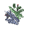
- Assembly
| Deposited unit | 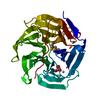
| ||||||||
|---|---|---|---|---|---|---|---|---|---|
| 1 |
| ||||||||
| Unit cell |
|
-Components
-Protein , 1 types, 1 molecules A
| #1: Protein | Mass: 34201.707 Da / Num. of mol.: 1 Source method: isolated from a genetically manipulated source Source: (gene. exp.) Photinus pyralis (common eastern firefly)Plasmid: pET15 / Production host:  |
|---|
-Non-polymers , 5 types, 217 molecules 


| #2: Chemical | ChemComp-HG /  Mass: 200.590 Da / Num. of mol.: 1 / Source method: obtained synthetically / Formula: Hg Mass: 200.590 Da / Num. of mol.: 1 / Source method: obtained synthetically / Formula: Hg | ||||
|---|---|---|---|---|---|
| #3: Chemical | ChemComp-MG /  Mass: 24.305 Da / Num. of mol.: 1 / Source method: obtained synthetically / Formula: Mg Mass: 24.305 Da / Num. of mol.: 1 / Source method: obtained synthetically / Formula: Mg | ||||
| #4: Chemical |  Mass: 118.174 Da / Num. of mol.: 2 / Source method: obtained synthetically / Formula: C6H14O2 / Comment: precipitant*YM Mass: 118.174 Da / Num. of mol.: 2 / Source method: obtained synthetically / Formula: C6H14O2 / Comment: precipitant*YM#5: Chemical | ChemComp-GOL / |  Mass: 92.094 Da / Num. of mol.: 1 / Source method: obtained synthetically / Formula: C3H8O3 Mass: 92.094 Da / Num. of mol.: 1 / Source method: obtained synthetically / Formula: C3H8O3#6: Water | ChemComp-HOH / | Mass: 18.015 Da / Num. of mol.: 212 / Source method: isolated from a natural source / Formula: H2O |
-Details
| Sequence details | Depositors state that the protein sequence should be the same as the database sequence UNP Q95YI4. ...Depositors state that the protein sequence should be the same as the database sequence UNP Q95YI4. The accession number is AB062786.2 for the sequence. |
|---|
-Experimental details
-Experiment
| Experiment | Method: X-RAY DIFFRACTION / Number of used crystals: 1 |
|---|
- Sample preparation
| Crystal | Density Matthews: 2.27 Å3/Da / Density % sol: 45.81 % Description: THE ENTRY CONTAINS FRIEDEL PAIRS IN F_PLUS/MINUS COLUMNS. |
|---|---|
| Crystal grow | Temperature: 293 K / Method: batch mode / pH: 7.5 Details: 150 uL of purified LRE solution (30 mg/ml LRE, 10 mM HEPES(pH7.5), 100 mM NaCl, 10 %(v/v) glycerol) was mixed with 240 uL of precipitant solution (31.3 %(w/v) PEG3350, 0.1 M HEPES(pH7.5), 10 ...Details: 150 uL of purified LRE solution (30 mg/ml LRE, 10 mM HEPES(pH7.5), 100 mM NaCl, 10 %(v/v) glycerol) was mixed with 240 uL of precipitant solution (31.3 %(w/v) PEG3350, 0.1 M HEPES(pH7.5), 10 %(v/v) MPD, 0.2 M MgCl2) and 15 uL of seed-crystal solution (31.3 %(w/v) PEG3350, 0.1 M HEPES(pH7.5), 10 %(v/v) MPD, 0.2 M MgCl2, 0.1 M NaCl, 5 %(v/v) glycerol) |
-Data collection
| Diffraction | Mean temperature: 100 K | ||||||||||||||||||||||||||||||||||||||||||||||||||||||||||||
|---|---|---|---|---|---|---|---|---|---|---|---|---|---|---|---|---|---|---|---|---|---|---|---|---|---|---|---|---|---|---|---|---|---|---|---|---|---|---|---|---|---|---|---|---|---|---|---|---|---|---|---|---|---|---|---|---|---|---|---|---|---|
| Diffraction source | Source: SYNCHROTRON / Site: SPring-8  / Beamline: BL41XU / Wavelength: 0.9839 Å / Beamline: BL41XU / Wavelength: 0.9839 Å | ||||||||||||||||||||||||||||||||||||||||||||||||||||||||||||
| Detector | Type: DECTRIS PILATUS3 6M / Detector: PIXEL / Date: May 30, 2016 | ||||||||||||||||||||||||||||||||||||||||||||||||||||||||||||
| Radiation | Monochromator: Si (111) / Protocol: SINGLE WAVELENGTH / Monochromatic (M) / Laue (L): M / Scattering type: x-ray | ||||||||||||||||||||||||||||||||||||||||||||||||||||||||||||
| Radiation wavelength | Wavelength: 0.9839 Å / Relative weight: 1 | ||||||||||||||||||||||||||||||||||||||||||||||||||||||||||||
| Reflection | Resolution: 1.6→50 Å / Num. obs: 79457 / % possible obs: 100 % / Redundancy: 27.894 % / CC1/2: 0.9904369 / Net I/σ(I): 4.144122 / Num. measured all: 2216376 | ||||||||||||||||||||||||||||||||||||||||||||||||||||||||||||
| Reflection shell |
|
- Processing
| Software |
| |||||||||||||||||||||||||||||||||||||||||||||||||||||||||||||||||||||||||||||||||||||||||||||||||||||||||||||||||||||||||||||||||||||||||||||||||||||||||||||||||
|---|---|---|---|---|---|---|---|---|---|---|---|---|---|---|---|---|---|---|---|---|---|---|---|---|---|---|---|---|---|---|---|---|---|---|---|---|---|---|---|---|---|---|---|---|---|---|---|---|---|---|---|---|---|---|---|---|---|---|---|---|---|---|---|---|---|---|---|---|---|---|---|---|---|---|---|---|---|---|---|---|---|---|---|---|---|---|---|---|---|---|---|---|---|---|---|---|---|---|---|---|---|---|---|---|---|---|---|---|---|---|---|---|---|---|---|---|---|---|---|---|---|---|---|---|---|---|---|---|---|---|---|---|---|---|---|---|---|---|---|---|---|---|---|---|---|---|---|---|---|---|---|---|---|---|---|---|---|---|---|---|---|---|
| Refinement | Method to determine structure: FOURIER SYNTHESIS Starting model: 5GTQ Resolution: 1.6→42.302 Å / SU ML: 0.29 / Cross valid method: FREE R-VALUE / σ(F): 1.33 / Phase error: 27.53 / Stereochemistry target values: ML Details: SF FILE CONTAINS FRIEDEL PAIRS UNDER I/F_MINUS AND I/F_PLUS COLUMNS.
| |||||||||||||||||||||||||||||||||||||||||||||||||||||||||||||||||||||||||||||||||||||||||||||||||||||||||||||||||||||||||||||||||||||||||||||||||||||||||||||||||
| Solvent computation | Shrinkage radii: 0.9 Å / VDW probe radii: 1.11 Å / Solvent model: FLAT BULK SOLVENT MODEL | |||||||||||||||||||||||||||||||||||||||||||||||||||||||||||||||||||||||||||||||||||||||||||||||||||||||||||||||||||||||||||||||||||||||||||||||||||||||||||||||||
| Displacement parameters | Biso max: 59.02 Å2 / Biso mean: 25.1018 Å2 / Biso min: 13.15 Å2 | |||||||||||||||||||||||||||||||||||||||||||||||||||||||||||||||||||||||||||||||||||||||||||||||||||||||||||||||||||||||||||||||||||||||||||||||||||||||||||||||||
| Refinement step | Cycle: final / Resolution: 1.6→42.302 Å
| |||||||||||||||||||||||||||||||||||||||||||||||||||||||||||||||||||||||||||||||||||||||||||||||||||||||||||||||||||||||||||||||||||||||||||||||||||||||||||||||||
| Refine LS restraints |
| |||||||||||||||||||||||||||||||||||||||||||||||||||||||||||||||||||||||||||||||||||||||||||||||||||||||||||||||||||||||||||||||||||||||||||||||||||||||||||||||||
| LS refinement shell | Refine-ID: X-RAY DIFFRACTION / Total num. of bins used: 22
|
