 Movie
Movie Controller

[English] 日本語
 Yorodumi
- PDB-5gvt: Crystal structures of the serine protease domain of murine plasma... -
+ Open data

- Basic information
| Entry | Database: PDB / ID: 5gvt | ||||||
|---|---|---|---|---|---|---|---|
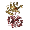
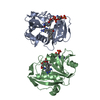
| Title | Crystal structures of the serine protease domain of murine plasma kallikrein | ||||||
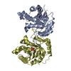
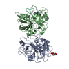
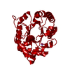
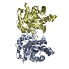
 Components Components | Plasma kallikrein | ||||||
 Keywords Keywords | HYDROLASE / serine protease / murine plasma kallikrein | ||||||
| Function / homology |  Function and homology information Function and homology informationIntrinsic Pathway of Fibrin Clot Formation / Activation of Matrix Metalloproteinases / plasma kallikrein / positive regulation of fibrinolysis / plasminogen activation / fibrinolysis / blood coagulation / inflammatory response / serine-type endopeptidase activity / extracellular space Similarity search - Function | ||||||
| Biological species |  | ||||||
| Method |  X-RAY DIFFRACTION / SYNCHROTRON / MOLECULAR REPLACEMENT / Resolution: 2.61 Å X-RAY DIFFRACTION / SYNCHROTRON / MOLECULAR REPLACEMENT / Resolution: 2.61 Å | ||||||
 Authors Authors | Xu, M. / Jiang, L. / Huang, M. | ||||||
| Funding support |  China, 1items China, 1items
| ||||||
 Citation Citation | Journal: Chin.J.Struct.Chem. / Year: 2017 Title: Crystal Structures of the Serine Protease Domain of Murine Plasma Kallikrein Authors: Xu, M. / Xu, P. / Zhou, X. / Andreasen, P. / Jiang, L. / Huang, M. | ||||||
| History |
|
- Structure visualization
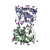
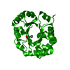
| Structure viewer | Molecule: MolmilJmol/JSmol |
|---|
- Downloads & links
-Download
| PDBx/mmCIF format | 5gvt.cif.gz | 105.4 KB | Display | PDBx/mmCIF format |
|---|---|---|---|---|
| PDB format | pdb5gvt.ent.gz | 79.5 KB | Display | PDB format |
| PDBx/mmJSON format | 5gvt.json.gz | Tree view | PDBx/mmJSON format | |
| Others |  Other downloads Other downloads |
-Validation report
| Arichive directory | https://data.pdbj.org/pub/pdb/validation_reports/gv/5gvtftp://data.pdbj.org/pub/pdb/validation_reports/gv/5gvt | HTTPS FTP |
|---|
-Related structure data
| Related structure data |  2anyS S: Starting model for refinement |
|---|---|
| Similar structure data |


-Links
 PDBj
PDBj


- Assembly
| Deposited unit | 
| ||||||||
|---|---|---|---|---|---|---|---|---|---|
| 1 |
| ||||||||
| Unit cell |
|
-Components
| #1: Protein | Mass: 27859.521 Da / Num. of mol.: 2 Fragment: Plasma kallikrein light chain, serine protease domain Mutation: C122S Source method: isolated from a genetically manipulated source Source: (gene. exp.)  Komagataella pastoris (fungus) / References: UniProt: P26262, plasma kallikrein Komagataella pastoris (fungus) / References: UniProt: P26262, plasma kallikrein#2: Sugar |   Type: D-saccharide, beta linking / Mass: 180.156 Da / Num. of mol.: 2 Type: D-saccharide, beta linking / Mass: 180.156 Da / Num. of mol.: 2Source method: isolated from a genetically manipulated source Formula: C6H12O6 #3: Sugar |   Type: D-saccharide, alpha linking / Mass: 180.156 Da / Num. of mol.: 2 Type: D-saccharide, alpha linking / Mass: 180.156 Da / Num. of mol.: 2Source method: isolated from a genetically manipulated source Formula: C6H12O6 #4: Water | ChemComp-HOH / |  Mass: 18.015 Da / Num. of mol.: 14 / Source method: isolated from a natural source / Formula: H2O Mass: 18.015 Da / Num. of mol.: 14 / Source method: isolated from a natural source / Formula: H2OHas protein modification | Y | |
|---|
-Experimental details
-Experiment
| Experiment | Method: X-RAY DIFFRACTION / Number of used crystals: 1 |
|---|
- Sample preparation
| Crystal | Density Matthews: 3.03 Å3/Da / Density % sol: 59.41 % |
|---|---|
| Crystal grow | Temperature: 298 K / Method: evaporation / pH: 8.5 / Details: PEG 3350 25%, 0.1M Tris-HCl, 0.1M NaCl |
-Data collection
| Diffraction | Mean temperature: 100 K |
|---|---|
| Diffraction source | Source: SYNCHROTRON / Site: SSRF / Beamline: BL17U / Wavelength: 0.979 Å |
| Detector | Type: ADSC QUANTUM 315r / Detector: CCD / Date: Jul 11, 2014 |
| Radiation | Protocol: SINGLE WAVELENGTH / Monochromatic (M) / Laue (L): M / Scattering type: x-ray |
| Radiation wavelength | Wavelength: 0.979 Å / Relative weight: 1 |
| Reflection | Resolution: 2.6→50 Å / Num. obs: 21266 / % possible obs: 98.4 % / Redundancy: 3.9 % / Net I/σ(I): 13.7 |
- Processing
| Software |
| ||||||||||||||||||||||||||||||||||||||||||||||||||||||||
|---|---|---|---|---|---|---|---|---|---|---|---|---|---|---|---|---|---|---|---|---|---|---|---|---|---|---|---|---|---|---|---|---|---|---|---|---|---|---|---|---|---|---|---|---|---|---|---|---|---|---|---|---|---|---|---|---|---|
| Refinement | Method to determine structure: MOLECULAR REPLACEMENT Starting model: 2ANY Resolution: 2.61→46.04 Å / SU ML: 0.3 / Cross valid method: THROUGHOUT / σ(F): 1.36 / Phase error: 28.66 / Stereochemistry target values: ML
| ||||||||||||||||||||||||||||||||||||||||||||||||||||||||
| Solvent computation | Shrinkage radii: 0.9 Å / VDW probe radii: 1.11 Å / Solvent model: FLAT BULK SOLVENT MODEL | ||||||||||||||||||||||||||||||||||||||||||||||||||||||||
| Refinement step | Cycle: LAST / Resolution: 2.61→46.04 Å
| ||||||||||||||||||||||||||||||||||||||||||||||||||||||||
| Refine LS restraints |
| ||||||||||||||||||||||||||||||||||||||||||||||||||||||||
| LS refinement shell |
|
