| Entry | Database: PDB / ID: 5gvs
|
|---|
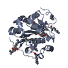
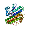
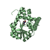
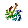
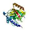
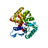
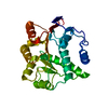
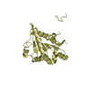
| Title | Crystal structure of the DDX41 DEAD domain in an apo open form |
|---|
 Components Components | Probable ATP-dependent RNA helicase DDX41 |
|---|
 Keywords Keywords | HYDROLASE / ATPase / DEAD box protein |
|---|
| Function / homology |  Function and homology information Function and homology information
STING mediated induction of host immune responses / IRF3-mediated induction of type I IFN / cGAS/STING signaling pathway / cellular response to interferon-beta / catalytic step 2 spliceosome / mRNA Splicing - Major Pathway / Regulation of innate immune responses to cytosolic DNA / spliceosomal complex / mRNA splicing, via spliceosome / defense response to virus ...STING mediated induction of host immune responses / IRF3-mediated induction of type I IFN / cGAS/STING signaling pathway / cellular response to interferon-beta / catalytic step 2 spliceosome / mRNA Splicing - Major Pathway / Regulation of innate immune responses to cytosolic DNA / spliceosomal complex / mRNA splicing, via spliceosome / defense response to virus / cell differentiation / RNA helicase activity / cell population proliferation / RNA helicase / mRNA binding / apoptotic process / endoplasmic reticulum / ATP hydrolysis activity / positive regulation of transcription by RNA polymerase II / DNA binding / RNA binding / zinc ion binding / nucleoplasm / ATP binding / membrane / nucleus / cytosolSimilarity search - Function DDX41, DEAD-box helicase domain / RNA helicase, DEAD-box type, Q motif / DEAD-box RNA helicase Q motif profile. / DEAD/DEAH box helicase domain / DEAD/DEAH box helicase / Helicase conserved C-terminal domain / helicase superfamily c-terminal domain / Superfamilies 1 and 2 helicase C-terminal domain profile. / Superfamilies 1 and 2 helicase ATP-binding type-1 domain profile. / DEAD-like helicases superfamily ...DDX41, DEAD-box helicase domain / RNA helicase, DEAD-box type, Q motif / DEAD-box RNA helicase Q motif profile. / DEAD/DEAH box helicase domain / DEAD/DEAH box helicase / Helicase conserved C-terminal domain / helicase superfamily c-terminal domain / Superfamilies 1 and 2 helicase C-terminal domain profile. / Superfamilies 1 and 2 helicase ATP-binding type-1 domain profile. / DEAD-like helicases superfamily / Helicase, C-terminal / Helicase superfamily 1/2, ATP-binding domain / P-loop containing nucleotide triphosphate hydrolases / Rossmann fold / P-loop containing nucleoside triphosphate hydrolase / 3-Layer(aba) Sandwich / Alpha BetaSimilarity search - Domain/homology |
|---|
| Biological species |  Homo sapiens (human) Homo sapiens (human) |
|---|
| Method |  X-RAY DIFFRACTION / SYNCHROTRON / MOLECULAR REPLACEMENT / Resolution: 2.2 Å X-RAY DIFFRACTION / SYNCHROTRON / MOLECULAR REPLACEMENT / Resolution: 2.2 Å |
|---|
 Authors Authors | Omura, H. / Oikawa, D. / Nakane, T. / Kato, M. / Ishii, R. / Goto, Y. / Suga, H. / Ishitani, R. / Tokunaga, F. / Nureki, O. |
|---|
| Funding support |  Japan, 1items Japan, 1items | Organization | Grant number | Country |
|---|
| | Japan |
|
|---|
 Citation Citation | Journal: Sci Rep / Year: 2016
Title: Structural and Functional Analysis of DDX41: a bispecific immune receptor for DNA and cyclic dinucleotide
Authors: Omura, H. / Oikawa, D. / Nakane, T. / Kato, M. / Ishii, R. / Ishitani, R. / Tokunaga, F. / Nureki, O. |
|---|
| History | | Deposition | Sep 6, 2016 | Deposition site: PDBJ / Processing site: PDBJ |
|---|
| Revision 1.0 | Oct 19, 2016 | Provider: repository / Type: Initial release |
|---|
| Revision 1.1 | Feb 26, 2020 | Group: Data collection / Derived calculations / Category: diffrn_source / pdbx_struct_oper_list
Item: _diffrn_source.pdbx_synchrotron_site / _pdbx_struct_oper_list.symmetry_operation |
|---|
| Revision 1.2 | Nov 8, 2023 | Group: Data collection / Database references / Refinement description
Category: chem_comp_atom / chem_comp_bond ...chem_comp_atom / chem_comp_bond / database_2 / pdbx_initial_refinement_model
Item: _database_2.pdbx_DOI / _database_2.pdbx_database_accession |
|---|
|
|---|
 Movie
Movie Controller


 Open data
 Basic information
 Structure visualization
 Downloads & links
 Other downloads


 PDBj
PDBj


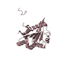
 Assembly


 Mass: 18.015 Da / Num. of mol.: 233 / Source method: isolated from a natural source / Formula: H2O
 Sample preparation
 Processing