 Movie
Movie Controller

[English] 日本語
 Yorodumi
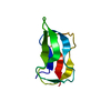
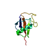
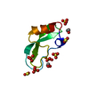
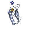
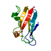
- PDB-5gua: Structure of biotin carboxyl carrier protein from pyrococcus hori... -
+ Open data

- Basic information
| Entry | Database: PDB / ID: 5gua | ||||||
|---|---|---|---|---|---|---|---|
| Title | Structure of biotin carboxyl carrier protein from pyrococcus horikoshi OT3 (delta N79) A138Y mutant | ||||||
 Components Components | 149aa long hypothetical methylmalonyl-CoA decarboxylase gamma chain | ||||||
 Keywords Keywords | TRANSFERASE / Surface engineering / Crystal packing / Crystal contact engineering | ||||||
| Function / homology |  Function and homology information Function and homology information: / Biotin-binding site / Biotin-requiring enzymes attachment site. / RNA polymerase II/Efflux pump adaptor protein, barrel-sandwich hybrid domain / Biotin-requiring enzyme / Biotinyl/lipoyl domain profile. / Biotin/lipoyl attachment / Single hybrid motif / OB fold (Dihydrolipoamide Acetyltransferase, E2P) / Beta Barrel / Mainly Beta Similarity search - Domain/homology | ||||||
| Biological species |   Pyrococcus horikoshii (archaea) Pyrococcus horikoshii (archaea) | ||||||
| Method |  X-RAY DIFFRACTION / SYNCHROTRON / MOLECULAR REPLACEMENT / Resolution: 1.5 Å X-RAY DIFFRACTION / SYNCHROTRON / MOLECULAR REPLACEMENT / Resolution: 1.5 Å | ||||||
 Authors Authors | Yamada, K. / Kunishima, N. / Matsuura, Y. / Nakai, K. / Naitow, H. / Fukasawa, Y. / Tomii, K. | ||||||
| Funding support |  Japan, 1items Japan, 1items
| ||||||
 Citation Citation | Journal: Acta Crystallogr D Struct Biol / Year: 2017 Title: Designing better diffracting crystals of biotin carboxyl carrier protein from Pyrococcus horikoshii by a mutation based on the crystal-packing propensity of amino acids. Authors: Yamada, K.D. / Kunishima, N. / Matsuura, Y. / Nakai, K. / Naitow, H. / Fukasawa, Y. / Tomii, K. | ||||||
| History |
|
- Structure visualization
| Structure viewer | Molecule: MolmilJmol/JSmol |
|---|
- Downloads & links
-Download
| PDBx/mmCIF format | 5gua.cif.gz | 28.8 KB | Display | PDBx/mmCIF format |
|---|---|---|---|---|
| PDB format | pdb5gua.ent.gz | 17.6 KB | Display | PDB format |
| PDBx/mmJSON format | 5gua.json.gz | Tree view | PDBx/mmJSON format | |
| Others |  Other downloads Other downloads |
-Validation report
| Arichive directory | https://data.pdbj.org/pub/pdb/validation_reports/gu/5guaftp://data.pdbj.org/pub/pdb/validation_reports/gu/5gua | HTTPS FTP |
|---|
-Related structure data
| Related structure data |  5gu8C  5gu9C 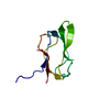 2evbS C: citing same article ( S: Starting model for refinement |
|---|---|
| Similar structure data |


-Links
 PDBj
PDBj

- Assembly
| Deposited unit | 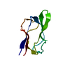
| |||||||||
|---|---|---|---|---|---|---|---|---|---|---|
| 1 |
| |||||||||
| Unit cell |
| |||||||||
| Components on special symmetry positions |
|
-Components
| #1: Protein | Mass: 7792.214 Da / Num. of mol.: 1 / Fragment: UNP RESIDUES 80-149 / Mutation: A138Y Source method: isolated from a genetically manipulated source Source: (gene. exp.) Pyrococcus horikoshii (strain ATCC 700860 / DSM 12428 / JCM 9974 / NBRC 100139 / OT-3) (archaea)Strain: ATCC 700860 / DSM 12428 / JCM 9974 / NBRC 100139 / OT-3 Gene: PH1284 / Plasmid: PET 11A / Production host:  |
|---|---|
| #2: Water | ChemComp-HOH /  Mass: 18.015 Da / Num. of mol.: 105 / Source method: isolated from a natural source / Formula: H2O Mass: 18.015 Da / Num. of mol.: 105 / Source method: isolated from a natural source / Formula: H2O |
-Experimental details
-Experiment
| Experiment | Method: X-RAY DIFFRACTION / Number of used crystals: 1 |
|---|
- Sample preparation
| Crystal | Density Matthews: 2.17 Å3/Da / Density % sol: 43.33 % |
|---|---|
| Crystal grow | Temperature: 293 K / Method: vapor diffusion, sitting drop / pH: 4.5 Details: 2.5M sodium chloride, 0.2M lithium sulfate, 0.1M acetate-NaOH |
-Data collection
| Diffraction | Mean temperature: 100 K |
|---|---|
| Diffraction source | Source: SYNCHROTRON / Site: SPring-8 / Beamline: BL26B2 / Wavelength: 1 Å |
| Detector | Type: MARMOSAIC 225 mm CCD / Detector: CCD / Date: Aug 3, 2015 |
| Radiation | Protocol: SINGLE WAVELENGTH / Monochromatic (M) / Laue (L): M / Scattering type: x-ray |
| Radiation wavelength | Wavelength: 1 Å / Relative weight: 1 |
| Reflection | Resolution: 1.5→33.8 Å / Num. obs: 11290 / % possible obs: 99.4 % / Redundancy: 9.9 % / Biso Wilson estimate: 10.4 Å2 / Rmerge(I) obs: 0.044 / Net I/σ(I): 41.2 |
| Reflection shell | Resolution: 1.5→1.53 Å / Redundancy: 6.1 % / Rmerge(I) obs: 0.102 / Mean I/σ(I) obs: 16 / % possible all: 98.2 |
- Processing
| Software |
| ||||||||||||||||||||
|---|---|---|---|---|---|---|---|---|---|---|---|---|---|---|---|---|---|---|---|---|---|
| Refinement | Method to determine structure: MOLECULAR REPLACEMENT Starting model: 2EVB Resolution: 1.5→33.8 Å / SU ML: 0.1 / Cross valid method: THROUGHOUT / Phase error: 21.34
| ||||||||||||||||||||
| Solvent computation | Shrinkage radii: 0.9 Å / VDW probe radii: 1.11 Å | ||||||||||||||||||||
| Displacement parameters | Biso mean: 17.8 Å2 | ||||||||||||||||||||
| Refinement step | Cycle: LAST / Resolution: 1.5→33.8 Å
| ||||||||||||||||||||
| LS refinement shell | Resolution: 1.5→1.65 Å
|
