 Movie
Movie Controller


+ Open data

- Basic information
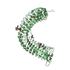
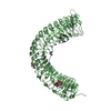
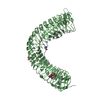
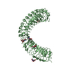
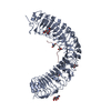
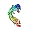
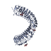
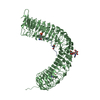
| Entry | Database: PDB / ID: 5gr9 | ||||||
|---|---|---|---|---|---|---|---|
| Title | Crystal structure of PXY-TDIF/CLE41 | ||||||
 Components Components |
| ||||||
 Keywords Keywords | TRANSFERASE / PXY / TDIF / CLE peptides / leucine rich repeat | ||||||
| Function / homology |  Function and homology information Function and homology informationprocambium histogenesis / phloem development / xylem development / cell-cell signaling involved in cell fate commitment / axillary shoot meristem initiation / maintenance of root meristem identity / phloem or xylem histogenesis / secondary shoot formation / apoplast / receptor serine/threonine kinase binding ...procambium histogenesis / phloem development / xylem development / cell-cell signaling involved in cell fate commitment / axillary shoot meristem initiation / maintenance of root meristem identity / phloem or xylem histogenesis / secondary shoot formation / apoplast / receptor serine/threonine kinase binding / regulation of cell differentiation / protein kinase activity / non-specific serine/threonine protein kinase / protein serine kinase activity / cell division / protein serine/threonine kinase activity / ATP binding / plasma membrane Similarity search - Function | ||||||
| Biological species |  | ||||||
| Method |  X-RAY DIFFRACTION / SYNCHROTRON / MOLECULAR REPLACEMENT / Resolution: 2.767 Å X-RAY DIFFRACTION / SYNCHROTRON / MOLECULAR REPLACEMENT / Resolution: 2.767 Å | ||||||
 Authors Authors | Chai, J.J. / Zhang, H.Q. | ||||||
 Citation Citation | Journal: Cell Res. / Year: 2016 Title: Crystal structure of PXY-TDIF complex reveals a conserved recognition mechanism among CLE peptide-receptor pairs Authors: Zhang, H.Q. / Lin, X.Y. / Han, Z.F. / Qu, L.J. / Chai, J.J. | ||||||
| History |
|
- Structure visualization
| Structure viewer | Molecule: MolmilJmol/JSmol |
|---|
- Downloads & links
-Download
| PDBx/mmCIF format | 5gr9.cif.gz | 134.8 KB | Display | PDBx/mmCIF format |
|---|---|---|---|---|
| PDB format | pdb5gr9.ent.gz | 102.9 KB | Display | PDB format |
| PDBx/mmJSON format | 5gr9.json.gz | Tree view | PDBx/mmJSON format | |
| Others |  Other downloads Other downloads |
-Validation report
| Arichive directory | https://data.pdbj.org/pub/pdb/validation_reports/gr/5gr9ftp://data.pdbj.org/pub/pdb/validation_reports/gr/5gr9 | HTTPS FTP |
|---|
-Related structure data
| Related structure data |  4mn8S S: Starting model for refinement |
|---|---|
| Similar structure data |


-Links
 PDBj
PDBj


- Assembly
| Deposited unit | 
| ||||||||
|---|---|---|---|---|---|---|---|---|---|
| 1 |
| ||||||||
| Unit cell |
|
-Components
| #1: Protein | Mass: 65982.461 Da / Num. of mol.: 1 / Fragment: UNP residues 31-629 Source method: isolated from a genetically manipulated source Source: (gene. exp.)   Spodoptera frugiperda (fall armyworm) Spodoptera frugiperda (fall armyworm)References: UniProt: Q9FII5, non-specific serine/threonine protein kinase | ||
|---|---|---|---|
| #2: Protein/peptide | Mass: 1280.322 Da / Num. of mol.: 1 / Source method: obtained synthetically / Source: (synth.) | ||
| #3: Sugar | ChemComp-NAG /   Type: D-saccharide, beta linking / Mass: 221.208 Da / Num. of mol.: 8 Type: D-saccharide, beta linking / Mass: 221.208 Da / Num. of mol.: 8Source method: isolated from a genetically manipulated source Formula: C8H15NO6 #4: Water | ChemComp-HOH / |  Mass: 18.015 Da / Num. of mol.: 36 / Source method: isolated from a natural source / Formula: H2O Mass: 18.015 Da / Num. of mol.: 36 / Source method: isolated from a natural source / Formula: H2O |
-Experimental details
-Experiment
| Experiment | Method: X-RAY DIFFRACTION / Number of used crystals: 1 |
|---|
- Sample preparation
| Crystal | Density Matthews: 3.75 Å3/Da / Density % sol: 67.21 % |
|---|---|
| Crystal grow | Temperature: 291 K / Method: vapor diffusion, hanging drop / pH: 5 Details: 16% w/v Polyethylene glycol 3350, 2% v/v Tacsimate pH 5.0, 0.1 M Sodium citrate tribasic dihydrate pH 5.5 PH range: 5.0-5.5 |
-Data collection
| Diffraction | Mean temperature: 100 K |
|---|---|
| Diffraction source | Source: SYNCHROTRON / Site: SSRF  / Beamline: BL17U / Wavelength: 0.979 Å / Beamline: BL17U / Wavelength: 0.979 Å |
| Detector | Type: RAYONIX MX-225 / Detector: CCD / Date: Mar 9, 2014 |
| Radiation | Protocol: SINGLE WAVELENGTH / Monochromatic (M) / Laue (L): M / Scattering type: x-ray |
| Radiation wavelength | Wavelength: 0.979 Å / Relative weight: 1 |
| Reflection | Resolution: 2.77→50 Å / Num. obs: 26768 / % possible obs: 99.7 % / Redundancy: 5.4 % / Net I/σ(I): 15.5 |
| Reflection shell | Resolution: 2.77→2.8 Å |
- Processing
| Software |
| |||||||||||||||||||||||||||||||||||||||||||||||||||||||||||||||||||||||||||||
|---|---|---|---|---|---|---|---|---|---|---|---|---|---|---|---|---|---|---|---|---|---|---|---|---|---|---|---|---|---|---|---|---|---|---|---|---|---|---|---|---|---|---|---|---|---|---|---|---|---|---|---|---|---|---|---|---|---|---|---|---|---|---|---|---|---|---|---|---|---|---|---|---|---|---|---|---|---|---|
| Refinement | Method to determine structure: MOLECULAR REPLACEMENT Starting model: 4MN8 Resolution: 2.767→39.905 Å / SU ML: 0.31 / Cross valid method: FREE R-VALUE / σ(F): 0 / Phase error: 24.18 / Stereochemistry target values: ML
| |||||||||||||||||||||||||||||||||||||||||||||||||||||||||||||||||||||||||||||
| Solvent computation | Shrinkage radii: 0.9 Å / VDW probe radii: 1.11 Å / Solvent model: FLAT BULK SOLVENT MODEL | |||||||||||||||||||||||||||||||||||||||||||||||||||||||||||||||||||||||||||||
| Refinement step | Cycle: LAST / Resolution: 2.767→39.905 Å
| |||||||||||||||||||||||||||||||||||||||||||||||||||||||||||||||||||||||||||||
| Refine LS restraints |
| |||||||||||||||||||||||||||||||||||||||||||||||||||||||||||||||||||||||||||||
| LS refinement shell |
|
