 Movie
Movie Controller

[English] 日本語
 Yorodumi
- PDB-5gkb: Crystal Structure of Fatty Acid-Binding Protein in Brain Tissue o... -
+ Open data

- Basic information
| Entry | Database: PDB / ID: 5gkb | ||||||
|---|---|---|---|---|---|---|---|
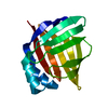
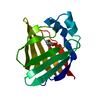
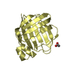
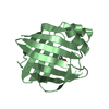
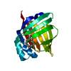
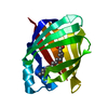
| Title | Crystal Structure of Fatty Acid-Binding Protein in Brain Tissue of Drosophila melanogaster without citrate inside | ||||||
 Components Components | Fatty acid bindin protein, isoform B | ||||||
 Keywords Keywords | LIPID BINDING PROTEIN / Fatty Acid Binding Protein | ||||||
| Function / homology |  Function and homology information Function and homology information | ||||||
| Biological species |  | ||||||
| Method |  X-RAY DIFFRACTION / SYNCHROTRON / MOLECULAR REPLACEMENT / Resolution: 2.04 Å X-RAY DIFFRACTION / SYNCHROTRON / MOLECULAR REPLACEMENT / Resolution: 2.04 Å | ||||||
 Authors Authors | Cheng, Y.-Y. / Huang, Y.-F. / Lin, H.-H. / Chang, W.W. / Lyu, P.-C. | ||||||
 Citation Citation | Journal: Biochim Biophys Acta Mol Cell Biol Lipids / Year: 2019 Title: The ligand-mediated affinity of brain-type fatty acid-binding protein for membranes determines the directionality of lipophilic cargo transport. Authors: Cheng, Y.-Y. / Huang, Y.-F. / Lin, H.-H. / Chang, W.W. / Lyu, P.-C. #1: Journal: To Be PublishedTitle: Revisit the Relationship of Thermal Stability and Purity in Recombinant Drosophila Fatty Acid-Binding protein Authors: Cheng, Y.-Y. / Huang, Y.-F. / Lin, H.-H. | ||||||
| History |
|
- Structure visualization
| Structure viewer | Molecule: MolmilJmol/JSmol |
|---|
- Downloads & links
-Download
| PDBx/mmCIF format | 5gkb.cif.gz | 63.1 KB | Display | PDBx/mmCIF format |
|---|---|---|---|---|
| PDB format | pdb5gkb.ent.gz | 47.2 KB | Display | PDB format |
| PDBx/mmJSON format | 5gkb.json.gz | Tree view | PDBx/mmJSON format | |
| Others |  Other downloads Other downloads |
-Validation report
| Arichive directory | https://data.pdbj.org/pub/pdb/validation_reports/gk/5gkbftp://data.pdbj.org/pub/pdb/validation_reports/gk/5gkb | HTTPS FTP |
|---|
-Related structure data
| Related structure data | 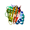 5ggeSC S: Starting model for refinement C: citing same article ( |
|---|---|
| Similar structure data |


-Links
 PDBj
PDBj

- Assembly
| Deposited unit | 
| ||||||||
|---|---|---|---|---|---|---|---|---|---|
| 1 |
| ||||||||
| Unit cell |
| ||||||||
| Components on special symmetry positions |
|
-Components
| #1: Protein | Mass: 14520.516 Da / Num. of mol.: 1 Source method: isolated from a genetically manipulated source Source: (gene. exp.)  |
|---|---|
| #2: Water | ChemComp-HOH /  Mass: 18.015 Da / Num. of mol.: 128 / Source method: isolated from a natural source / Formula: H2O Mass: 18.015 Da / Num. of mol.: 128 / Source method: isolated from a natural source / Formula: H2O |
-Experimental details
-Experiment
| Experiment | Method: X-RAY DIFFRACTION / Number of used crystals: 1 |
|---|
- Sample preparation
| Crystal | Density Matthews: 2.24 Å3/Da / Density % sol: 45.11 % |
|---|---|
| Crystal grow | Temperature: 293 K / Method: vapor diffusion, sitting drop / pH: 5.2 / Details: 0.1M phosphate citrate, 1.6M NaH2PO4, 0.4M K2HPO4 |
-Data collection
| Diffraction | Mean temperature: 110 K |
|---|---|
| Diffraction source | Source: SYNCHROTRON / Site: NSRRC  / Beamline: BL13C1 / Wavelength: 0.97622 Å / Beamline: BL13C1 / Wavelength: 0.97622 Å |
| Detector | Type: RAYONIX MX300HE / Detector: CCD / Date: Apr 12, 2015 |
| Radiation | Protocol: SINGLE WAVELENGTH / Monochromatic (M) / Laue (L): M / Scattering type: x-ray |
| Radiation wavelength | Wavelength: 0.97622 Å / Relative weight: 1 |
| Reflection | Resolution: 2.04→30 Å / Num. obs: 8811 / % possible obs: 99.98 % / Observed criterion σ(F): 0 / Observed criterion σ(I): -3 / Redundancy: 9.3 % / Rmerge(I) obs: 0.1 / Rsym value: 0.1 / Net I/σ(I): 22.6 |
| Reflection shell | Resolution: 2.04→2.11 Å / Redundancy: 9.1 % / Rmerge(I) obs: 0.433 / Mean I/σ(I) obs: 6.5 / % possible all: 99.9 |
- Processing
| Software |
| |||||||||||||||||||||||||||||||||||||||||||||||||
|---|---|---|---|---|---|---|---|---|---|---|---|---|---|---|---|---|---|---|---|---|---|---|---|---|---|---|---|---|---|---|---|---|---|---|---|---|---|---|---|---|---|---|---|---|---|---|---|---|---|---|
| Refinement | Method to determine structure: MOLECULAR REPLACEMENT Starting model: 5GGE Resolution: 2.04→28.091 Å / SU ML: 0.21 / Cross valid method: FREE R-VALUE / σ(F): 1.35 / Phase error: 22.91
| |||||||||||||||||||||||||||||||||||||||||||||||||
| Solvent computation | Shrinkage radii: 0.9 Å / VDW probe radii: 1.11 Å | |||||||||||||||||||||||||||||||||||||||||||||||||
| Refinement step | Cycle: LAST / Resolution: 2.04→28.091 Å
| |||||||||||||||||||||||||||||||||||||||||||||||||
| Refine LS restraints |
| |||||||||||||||||||||||||||||||||||||||||||||||||
| LS refinement shell |
|
