 Movie
Movie Controller

[English] 日本語
 Yorodumi
- PDB-5g5d: Crystal Structure of the CohScaC2-XDocCipA type II complex from C... -
+ Open data

- Basic information
| Entry | Database: PDB / ID: 5g5d | ||||||
|---|---|---|---|---|---|---|---|
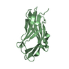
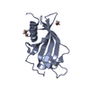
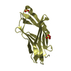
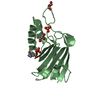
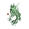
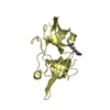
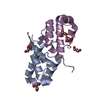
| Title | Crystal Structure of the CohScaC2-XDocCipA type II complex from Clostridium thermocellum | ||||||
 Components Components |
| ||||||
 Keywords Keywords | CARBOHYDRATE BINDING PROTEIN / COHESIN-DOCKERIN COMPLEX / TYPE II INTERACTION / SCAFFOLDIN / CELLULOSOME / C. THERMOCELLUM | ||||||
| Function / homology |  Function and homology information Function and homology informationS-layer / cellulose binding / polysaccharide catabolic process / cellulose catabolic process / hydrolase activity, hydrolyzing O-glycosyl compounds / cell wall organization / carbohydrate binding / extracellular region Similarity search - Function | ||||||
| Biological species |  RUMINICLOSTRIDIUM THERMOCELLUM AD2 (bacteria)CLOSTRIDIUM THERMOCELLUM (bacteria) RUMINICLOSTRIDIUM THERMOCELLUM AD2 (bacteria)CLOSTRIDIUM THERMOCELLUM (bacteria) | ||||||
| Method |  X-RAY DIFFRACTION / SYNCHROTRON / MOLECULAR REPLACEMENT / Resolution: 3 Å X-RAY DIFFRACTION / SYNCHROTRON / MOLECULAR REPLACEMENT / Resolution: 3 Å | ||||||
 Authors Authors | Carvalho, A.L. / A Bras, J.L. / Najmudin, S.H. / Pinheiro, B.A. / Fontes, C.M.G.A. | ||||||
 Citation Citation | Journal: Sci Rep / Year: 2016 Title: Diverse specificity of cellulosome attachment to the bacterial cell surface. Authors: Bras, J.L. / Pinheiro, B.A. / Cameron, K. / Cuskin, F. / Viegas, A. / Najmudin, S. / Bule, P. / Pires, V.M. / Romao, M.J. / Bayer, E.A. / Spencer, H.L. / Smith, S. / Gilbert, H.J. / Alves, V. ...Authors: Bras, J.L. / Pinheiro, B.A. / Cameron, K. / Cuskin, F. / Viegas, A. / Najmudin, S. / Bule, P. / Pires, V.M. / Romao, M.J. / Bayer, E.A. / Spencer, H.L. / Smith, S. / Gilbert, H.J. / Alves, V.D. / Carvalho, A.L. / Fontes, C.M. | ||||||
| History |
|
- Structure visualization
| Structure viewer | Molecule: MolmilJmol/JSmol |
|---|
- Downloads & links
-Download
| PDBx/mmCIF format | 5g5d.cif.gz | 132.8 KB | Display | PDBx/mmCIF format |
|---|---|---|---|---|
| PDB format | pdb5g5d.ent.gz | 105.4 KB | Display | PDB format |
| PDBx/mmJSON format | 5g5d.json.gz | Tree view | PDBx/mmJSON format | |
| Others |  Other downloads Other downloads |
-Validation report
| Summary document | 5g5d_validation.pdf.gz | 440.4 KB | Display | wwPDB validaton report |
|---|---|---|---|---|
| Full document | 5g5d_full_validation.pdf.gz | 442.1 KB | Display | |
| Data in XML | 5g5d_validation.xml.gz | 12.9 KB | Display | |
| Data in CIF | 5g5d_validation.cif.gz | 16.6 KB | Display | |
| Arichive directory | https://data.pdbj.org/pub/pdb/validation_reports/g5/5g5dftp://data.pdbj.org/pub/pdb/validation_reports/g5/5g5d | HTTPS FTP |
-Related structure data
| Related structure data |  5k39C 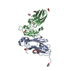 5m0yC  2bm3S S: Starting model for refinement C: citing same article ( |
|---|---|
| Similar structure data |


-Links
 PDBj
PDBj
- Assembly
| Deposited unit | 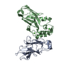
| ||||||||
|---|---|---|---|---|---|---|---|---|---|
| 1 | 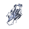
| ||||||||
| 2 | 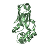
| ||||||||
| Unit cell |
|
-Components
| #1: Protein | Mass: 18947.541 Da / Num. of mol.: 1 / Fragment: SECOND COHESIN DOMAIN Source method: isolated from a genetically manipulated source Source: (gene. exp.) RUMINICLOSTRIDIUM THERMOCELLUM AD2 (bacteria)Production host: | ||
|---|---|---|---|
| #2: Protein | Mass: 17779.150 Da / Num. of mol.: 1 / Fragment: DOCKERIN DOMAIN Source method: isolated from a genetically manipulated source Source: (gene. exp.) CLOSTRIDIUM THERMOCELLUM (bacteria) / Production host: | ||
| #3: Chemical |   Mass: 40.078 Da / Num. of mol.: 2 / Source method: obtained synthetically / Formula: Ca Mass: 40.078 Da / Num. of mol.: 2 / Source method: obtained synthetically / Formula: CaSequence details | THE FIRST 3 RESIDUES WERE INTRODUCED FROM THE VECTOR. THE FIRST 4 AND LAST FIVE RESIDUES ARE NOT ...THE FIRST 3 RESIDUES WERE INTRODUCED | |
-Experimental details
-Experiment
| Experiment | Method: X-RAY DIFFRACTION / Number of used crystals: 1 |
|---|
- Sample preparation
| Crystal | Density Matthews: 3.1 Å3/Da / Density % sol: 60 % / Description: NONE |
|---|---|
| Crystal grow | Details: 0.2M CACL2 0.1M HEPES 7.5 25% PEG4K |
-Data collection
| Diffraction | Mean temperature: 100 K |
|---|---|
| Diffraction source | Source: SYNCHROTRON / Site: ESRF  / Beamline: ID29 / Wavelength: 0.95372 / Beamline: ID29 / Wavelength: 0.95372 |
| Detector | Type: PILATUS / Detector: PIXEL / Date: Jun 21, 2013 |
| Radiation | Protocol: SINGLE WAVELENGTH / Monochromatic (M) / Laue (L): M / Scattering type: x-ray |
| Radiation wavelength | Wavelength: 0.95372 Å / Relative weight: 1 |
| Reflection | Resolution: 2.89→90.57 Å / Num. obs: 10000 / % possible obs: 99.1 % / Observed criterion σ(I): 2 / Redundancy: 1.9 % / Rmerge(I) obs: 0.19 / Net I/σ(I): 5.9 |
| Reflection shell | Resolution: 3→3.18 Å / Redundancy: 7.2 % / Rmerge(I) obs: 1.18 / Mean I/σ(I) obs: 1.5 / % possible all: 99 |
- Processing
| Software |
| ||||||||||||||||||||||||||||||||||||||||||||||||||||||||||||||||||||||||||||||||||||||||||||||||||||||||||||||||||||||||||||||||||||||||||||||||||||||||||||||||||||||||||||||||||||||
|---|---|---|---|---|---|---|---|---|---|---|---|---|---|---|---|---|---|---|---|---|---|---|---|---|---|---|---|---|---|---|---|---|---|---|---|---|---|---|---|---|---|---|---|---|---|---|---|---|---|---|---|---|---|---|---|---|---|---|---|---|---|---|---|---|---|---|---|---|---|---|---|---|---|---|---|---|---|---|---|---|---|---|---|---|---|---|---|---|---|---|---|---|---|---|---|---|---|---|---|---|---|---|---|---|---|---|---|---|---|---|---|---|---|---|---|---|---|---|---|---|---|---|---|---|---|---|---|---|---|---|---|---|---|---|---|---|---|---|---|---|---|---|---|---|---|---|---|---|---|---|---|---|---|---|---|---|---|---|---|---|---|---|---|---|---|---|---|---|---|---|---|---|---|---|---|---|---|---|---|---|---|---|---|
| Refinement | Method to determine structure: MOLECULAR REPLACEMENT Starting model: PDB ENTRY 2BM3 Resolution: 3→90.56 Å / Cor.coef. Fo:Fc: 0.908 / Cor.coef. Fo:Fc free: 0.87 / SU B: 62.591 / SU ML: 0.488 / Cross valid method: THROUGHOUT / ESU R Free: 0.523 / Stereochemistry target values: MAXIMUM LIKELIHOOD / Details: HYDROGENS HAVE BEEN ADDED IN THE RIDING POSITIONS
| ||||||||||||||||||||||||||||||||||||||||||||||||||||||||||||||||||||||||||||||||||||||||||||||||||||||||||||||||||||||||||||||||||||||||||||||||||||||||||||||||||||||||||||||||||||||
| Solvent computation | Ion probe radii: 0.8 Å / Shrinkage radii: 0.8 Å / VDW probe radii: 1.1 Å / Solvent model: MASK | ||||||||||||||||||||||||||||||||||||||||||||||||||||||||||||||||||||||||||||||||||||||||||||||||||||||||||||||||||||||||||||||||||||||||||||||||||||||||||||||||||||||||||||||||||||||
| Refinement step | Cycle: LAST / Resolution: 3→90.56 Å
| ||||||||||||||||||||||||||||||||||||||||||||||||||||||||||||||||||||||||||||||||||||||||||||||||||||||||||||||||||||||||||||||||||||||||||||||||||||||||||||||||||||||||||||||||||||||
| Refine LS restraints |
|